

The ability to noninvasively access metabolic processes during the visual cycle is crucial for developing therapies against retinal degenerative diseases. We have developed a protocol for obtaining in vivo two-photon excited fluorescence images of the fundus in the human eye.
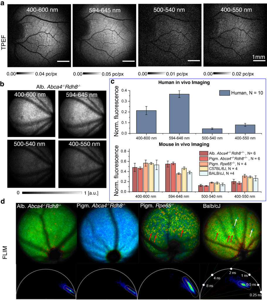
The visual cycle is a series of chemical transformations during which various fluorescent intermediates are formed. Blue-induced fundus autofluorescence allows visualization of retinal fluorophores. However, due to fundamental limitations, this method is limited to imaging lipofuscin, which contains byproducts of the visual cycle but does not directly reflect changes in photoreceptor function. The absorption spectra of fluorophores participating in the visual cycle, such as retinyl esters, lie in the UV spectral range. However, absorption and scattering in the front of the eye, as well as safety concerns, exclude UV from applications in ophthalmic imaging.
Two-photon excitation allows us to bypass this limitation and excite previously unavailable fluorophores, creating novel diagnostic capabilities. Retinal fluorophores can be excited with minimal absorption and much lower scattering and phototoxicity using femtosecond pulses at near-infrared wavelengths. Previously, we have shown imaging with a two-photon excited fluorescence scanning laser ophthalmoscope (TPEF-SLO) proved to be very useful in mice. Recently, in 2022 we report for the first time in vivo imaging of the human eye using a two-photon excited fluorescence (TPEF) with near-infrared light. We have built a compact instrument based on scanning laser ophthalmoscope (SLO) with a custom femtosecond fiber-based laser and efficient photon detection, and designed advanced data post-processing that enabled measurement of TPEF signals on the sub-single photon level. As a result, we can visualize the distribution of retinal fluorophores with exposure far below the safety limits for the human eye.
We heave measured dozens of volunteers to confirm the robustness of the technique and extend the experimental setup to work with mice models to investigate different eye diseases, like age-related or Stargardt macular degeneration. Imaging was performed in a dark room with no prior dark adaptation or pupil dilation. We were able to record the fluorescence signal and reconstruct the image in the majority of cases. This technique allays safety concerns by allowing for the acquisition of informative images at low laser exposure. We confirmed the applicability of the system for future clinical use of this imaging modality. These results constitute an essential step towards functional imaging of the human eye that directly reflects local changes in the retina’s function.
Author: Michał Dąbrowski, PhD
Team:
Publications:
- G. Palczewska, J. Boguslawski, P. Stremplewski, Ł. Kornaszewski, J. Zhang, Z. Dong, X.-X. Liang, E. Gratton, A. Vogel, M. Wojtkowski, and K. Palczewski, Noninvasive two-photon optical biopsy of retinal fluorophores, Proc. Natl. Acad. Sci. U. S. A 117, 22532 (2020)
- D. Stachowiak, J. Bogusławski, A. Głuszek, Z. Łaszczych, M. Wojtkowski, and G. Soboń, “Frequency-doubled femtosecond Er-doped fiber laser for two-photon excited fluorescence imaging, Biomed. Opt. Express 11, 4431 (2020)
- J. Boguslawski, G. Palczewska, S. Tomczewski, J. Milkiewicz, P.Kasprzycki, D. Stachowiak, K. Komar, M. J. Marzejon, B. L. Sikorski, A. Hudzikowski, A. Głuszek, Z. Łaszczych, K. Karnowski, G. Soboń, K. Palczewski, and M. Wojtkowski, In vivo imaginh of the human eye using a 2-photon-excited fluorescence scanning laser ophthalmoscope, JCI 132, 154218 (2022)
- J. Bogusławski, S. Tomczewski, M. Dąbrowski, K. Komar, J. Milkiewicz, G. Palczewska, K. Palczewski, and M. Wojtkowski, In vivo imaging of the muna retina using a two-photon excited fluorescence ophthalmoscope, STAR Protocols 4, 102225 (2023)
- G. Palczewska, M. Wojtkowski, and K. Palczewski, From mouse to human: Accessing the biochemistry of vision in vivo by two-photon excitation, Prog. Retin. Eye Res. 93, 101170 (2023)


