


Thanks to medical progress, we can cure more and more vision-related diseases and the bottleneck of successful ophthalmological interventions is diagnostics. The stage at which changes in the retina are detected directly translates into the patient’s chances of recovery. One of the most innovative and fastest-growing ophthalmological techniques is optoretinography (ORG), the leader of which in Poland is Prof. Maciej Wojtkowski from ICTER. Many centers around the world research ORG, and many use the treasure trove of knowledge of ICTER scientists.
One of the most important research centers specializing in ORG in the United States is the University of California at Davis (UC Davis), where Prof. Robert Zawadzki – a graduate of the Nicolaus Copernicus University in Toruń and a long-time collaborator of ICTER – has worked for about 20 years. We asked him what he does at UC Davis; how his research can translate into patients’ health; what are his feelings after visiting ICTER and why cooperation between centers from all over the world is crucial for the future of ophthalmology.
Please tell us what project you are working on and with whom during your current visit to ICTER.
Yes, this is my follow-up visit to ICTER at the invitation of Prof. M. Wojtkowski. Our plan during my previous visit was to assist in two research projects. One involved setting up and testing a fundus camera, which was designed for the STOC-T system and is now used for functional eye imaging. This research was conducted in collaboration with Dr. Andrea Curatolo’s team, with Wiktor Kulesza and Piotr Węgrzyn. During that stay, we managed to obtain an image of the mouse eye fundus using the camera, which could be used to determine the precise location of the retina for subsequent functional measurements using STOC-T. The second project involved cooperation with Dr. Michał Dąbrowski, that focused on assisting in the two-photon fluorescence imaging of the retina. Here, I also helped Mr. Michał correct the image from the auxiliary single-photon scanning light ophthalmoscope system, for two-photon measurements. In both the STOC-T and two-photon projects, the primary scientific instruments did not have real-time high-quality images, and this is where assistance was needed to build systems that would help align the eyes during the examination. During my current stay, I also participated in the CRATER conference and then focused mainly on working with Dr. Andrea Curatolo’s team. We collaborated on a manuscript describing the STOC-T measurement system for mouse imaging and its applications. Additionally, we discussed issues related to finding permissible light exposure limits for STOC-T measurements on experimental animals, as well as the details of the STOC-T optical system and the potential impact of various components on the measurement system’s resolution.
This is not the first time you have come to our centre. Please tell us briefly about what has been achieved during your previous visits and in what direction further cooperation with ICTER researchers is going.
Indeed, these visits are a continuation of my previous ones. I have previously collaborated with the same teams. During one of my first visits, taking place over two years ago, Dr. Michał Dąbrowski was still building his system, so our collaboration was limited to assisting in selecting certain optical components, which were needed for the experiments we conducted in 2022. In the case of Dr. Andrea Curatolo, during my first visits, we collaborated on the design, construction, and initial setup of the system. I hope that both projects will continue to develop and, in the case of Dr. Curatolo, allow for functional measurements at the retina in laboratory animals using STOC-T, and in the case of Dr. Dąbrowski, be useful for two-photon fluorescence measurements that may provide us with more information about diseases of photoreceptors and retinal pigment epithelium, cells containing majority of fluorescent molecules in the eye.
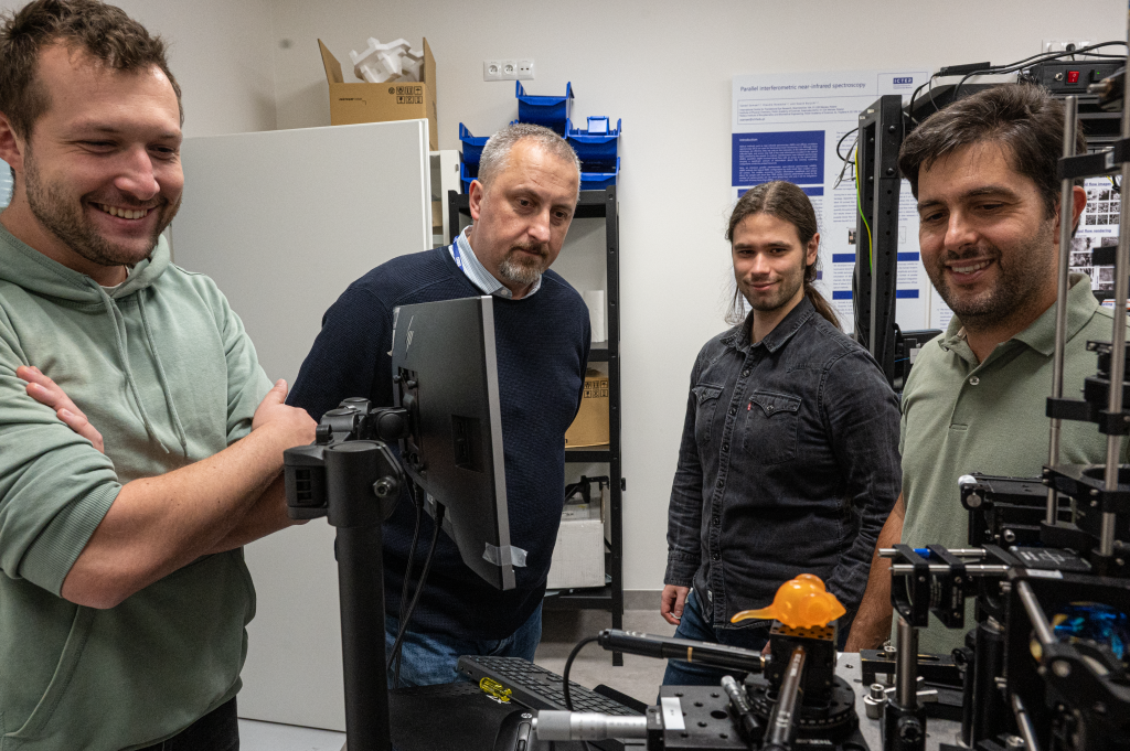
From the left: Piotr Węgrzyn, Prof. Robert Zawadzki, Wiktor Kulesza and Dr. Andrea Curatolo at ICTER’s lab.
What areas do you specialize in and what are the unique effects of this non-obvious combination in practice?
I specialize in the field of biomedical engineering or bio-photonics, specifically focusing on building and utilizing systems for functional measurements in the eye, particularly on the retina, in both humans and experimental animals. The outcomes of my work involve the development of new devices that enable the measurement of functional changes at the cellular level resulting from disease-related changes or age-related alterations. In the future, these methods may contribute to better diagnostics and the assessment of the effectiveness of gene or stem cell therapies in such cases.
Please tell us about your research and work at UC Davis.
I have been at UC Davis for about 20 years now, and currently, I’m a professor in the Department of Ophthalmology & Vision Science. I’m a member of two research groups there. One group is focused on testing and building clinical research devices; it’s called CHOIR, which stands for the Centre for Human Ophthalmic Imaging Research. The other research group I’m part of, EyePod Small Animal Ocular Imaging Laboratory, is involved in small animal ocular imaging. We design and create new devices for structural and functional eye measurements, mainly in mice and small experimental animals. The research we conduct aims to develop new methods that can be useful for both clinical doctors and scientists working on fundamental medical research, where new methods such as gene and stem cell therapy are being developed. We are one of the groups that helps other research teams test their innovations more effectively and identify potential issues faster while also aiding them in discovering new directions for the development of these various therapies.
How can you translate your research results into measurable and useful applications for patients?
Our research has the potential to be useful for patients in the following two scenarios. The first is the development of devices to enhance the diagnosis of eye diseases, improving these methods to the extent that even for individuals who do not yet exhibit any objective changes in their vision, it will be possible to determine whether there are any underlying changes. This is particularly crucial among individuals with genetic predispositions that put them at an increased risk. Knowing a person’s specific genetic defect can help tailor diagnostic methods to identify functional changes in certain cells, potentially allowing for prevention or at least a delay in the progression of the disease, given the current state of the medicine. In cases where these therapies are expensive, this is indeed a significant aspect. The second direction of our research is to confirm whether the methods used to treat patients are effective. In this scenario, if we find no changes, the doctor may choose another method that produces better results. This aspect is perhaps more tangible for patients. Of course, our research is also crucial in the implementation of new therapeutic methods as it accelerates the development of these therapies.
Please tell us how the optoretinography technology developed with ICTER is state-of-the-art, where in the world is it currently being developed, and what is your unique contribution to its development?
The technology of optoretinography, referred to as the method of measuring the functional response of the retina to light stimulation, has unique diagnostic potential and is, therefore one of a kind, it is currently being researched in various laboratories to understand how the signals measured using ORG can be linked to known physiological functions of individual retinal neurons. In Europe is primarily being developed by groups like ICTER led by Maciej Wojtkowski, a strong group in Germany under Geron Huettmann, and another group in Paris, lead by Kate Grieve. In United States, we have groups at UC Davis, University of Washington led by Ramkumar Sabesan, Indiana University led by Don Miller. There are also groups at the University of Illinois Chicago, the University of Wisconsin, the University of Pennsylvania and at Stanford University, just to name a few. All these groups focus on various aspects of ORG. My contribution to the development of this method involves e elucidating the physiological factors related to these signals. We have been able to confirm that changes in retinal water content are responsible for a portion of the signals we measure. This is a secondary effect following photoreceptor light activation but is related to water. Additionally, in our group, which develops devices for clinical research, we aim to create models that would enable us to validate our results, making it easier to determine the main characteristics of the optoretinography signal. Our research is directed towards finding better methods for optoretinographic measurements, discovering what truly influences the signal we measure. Note that we mainly measure changes in the thickness of certain retinal layers and alterations in light scattering. We are also developing methods to model these signals and more easily find correlations between the parameters of these curves and various eye diseases.
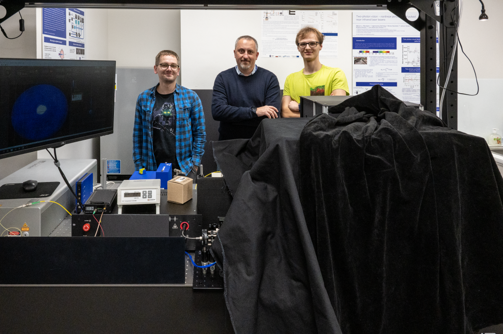
From the left: Dr. Michał Dąbrowski, Prof. Robert Zawadzki, and Bartłomiej Bałamut at ICTER’s lab.
Please specify how your career and approach to science have been influenced by the different locations and units where you have worked so far: undergraduate and graduate studies at UMK in Toruń, PhD in Vienna, and work at UC Davis.
My undergrad studies at Nicholaus Copernicus University (UMK) in Toruń were indeed essential for me to find myself where I am now, but, as in most cases, one’s career path and life journey are highly individual and challenging to replicate for others. They are often the result of certain coincidences and opportunities that have appeared on my path, some of which I was able to seize while others eluded me. However, my undergraduate studies at UMK were crucial in gaining fundamental knowledge in experimental physics and the use of computers in physics. They laid the foundation for my understanding of the scientific alphabet, so to speak. Then, during my master’s studies, I had the incredible fortune to start collaborating with Prof. Andrzej Kowalczyk, who, in the late 1990s, had a European Tempus grant for sending young students on various internships. In my case, I had the opportunity to intern at a university in Vienna, where I first encountered the method, I currently work on, Optical Coherence Tomography (OCT), and I also met one of its inventors, Prof. Adolf Fercher. After completing my master’s degree, I received an offer to pursue a Ph.D. in Vienna under Prof. Fercher, which is when I embarked on my doctoral studies. This experience allowed me to become proficient in both the OCT method and the field of biophotonics, as well as understand how to design devices for studying the eyes and other organs and how to apply data analysis methods. Therefore, my doctoral work was instrumental in building the knowledge needed for what I do now. After completing my Ph.D. in Vienna, I worked briefly as an assistant at UMK, and then, after about six months, I received a job offer at UC Davis as a Postdoc in John Werner Laboratory. It was there that I engaged in a significant project funded by the National Eye Institute, which involved building the world’s first system that combined adaptive optics with OCT. The knowledge I had gained during my doctoral studies, particularly in using OCT to study corneal shape and detect eye aberrations, proved to be ideal for this project, as I already had a foundation in ocular aberrations and understanding the function of the eye as an imaging element, as well as the basics of OCT. This experience in Vienna had also allowed me to become familiar with then up-and-coming detection technique known as Fourier Domain OCT. So, when I went to UC Davis, I had all the knowledge necessary to complete this project, which involved creating the first working adaptive optics OCT (AO-OCT) system, and we demonstrated the first images with cellular resolution on the retina. Throughout the years working at UC Davis, I maintained collaborations with groups in Toruń, led by Prof. Maciej Wojtkowski, and in Vienna. As these eye imaging methods evolved, we contributed to their development, primarily focusing on optical coherence tomography angiography, a method for non-invasively measuring blood flow in the eye. We also worked on methods that combined several different imaging techniques such as OCT with SLO, which are utilized by many modern systems for retinal eye imaging. About 12 years ago I began using these systems for measurements in experimental animals. This was made possible through collaboration between UC Davis Department of Ophthalmology (Prof. John Werner’s group) and the Department of Physiology, where Prof. Edward Pugh was involved. Through our collaboration with Ed Pugh, we created the EyePod team, which focused on studying the retina in experimental animals. It was around 2015 when we began working on ORG, or optoretinography. Thus, I continue to work in the same field, which I have been engaged in since my master’s studies, namely development and application of OCT in Medicine. I was able to do it by constantly applying the latest research method and technology. I have also been able to continuously expand my knowledge to keep my work as interesting and attractive as possible for these new emerging application fields.
What would you like to pass on to fellow scientists involved in eye research and the development of new ophthalmic therapies?
I would like to say that despite the fact that our new research methods and therapies seem very advanced, there are still many things we don’t know and cannot measure yet. I suspect that there is still a lot of work ahead of us to make these methods we are working on clinically available. Just as all these fields are still evolving, I would recommend young scientists to look at the current issues related to eye research. Perhaps even their individual experiences can be crucial in finding further solutions. So, the development of novel structural and functional assessment of the eye is something worth continuously engaging with.
Thank you very much for this interview, Prof. Robert Zawadzki. We eagerly anticipate further fruitful collaboration in the future.
Special thanks to all the ICTER scientists who participated in the photo session at our laboratories.
The interview was conducted by Dr. Anna Przybyło-Józefowicz (September 2023)
Title, introduction, and social media material: Journalist Marcin Powęska
Pictures: Dr. Karol Karnowski


