Retinal pigment epithelium (RPE) located at the back of the eye is essential for vision. It supports the photoreceptors, providing molecules required for their function. One of the main proteins produced by the RPE and indispensable for vision is the RPE65 enzyme, which is responsible for chemical signaling at the initial step of visual processing. De novo nonsense mutations in the Rpe65 gene underlie inherited genetic disorders of the eyes, resulting in blindness. To address this problem, we have harnessed the power of adenine base editors (ABEs) with Cas9 – single-guide RNA machinery to target the mutations in the Rpe65 gene for their repair. We delivered genes coding for ABEs and the Cas9 system subretinally via a lentiviral vector. Our therapeutic manipulation corrected the pathogenic mutation in a mouse model with up to 29% efficiency and with minimal formation of indel and off-target mutations. The ABE-treated mice displayed restored RPE65 expression and its activity in the visual cycle. Moreover, we have observed near-normal levels of retinal and visual functions. Our findings motivate the further testing of ABEs for the treatment of inherited retinal diseases and for the correction of pathological mutations with non-canonical protospacer-adjacent motifs.
Authors:
dr Andrzej Foik, e-mail: afoik@ichf.edu.pl & dr Anna Posłuszny, e-mail: aposluszny@ichf.edu.pl
Pertinent published article:
Restoration of visual function in adult mice with an inherited retinal disease via adenine base editing
Susie Suh, Elliot H. Choi, Henri Leinonen, Andrzej T. Foik, Gregory A. Newby, Wei-Hsi Yeh, Zhiqian Dong, Philip D. Kiser, David C. Lyon, David R. Liu & Krzysztof Palczewski, Nat Biomed Eng. 2021 Feb;5(2):169-178.
The retina is an essential part of the eye, as it is responsible for transforming light into electrical signals, which are processed in the brain later on. It effectively works as a photodetector of the eye. Imaging the structure and function of the living retina is crucial for the effective diagnosis and treatment of eye diseases and drug developments. Nowadays, structural information about the retina can be obtained from, e.g., OCT studies. However, functional alterations are the first signs of early pathological processes and often precede structural changes; this information is currently very challenging to obtain.
The retina has a layered structure, which is packed with various fluorophores. For example, retinal pigments epithelium (RPE) contains lipofuscin, a byproduct of the visual cycle. Lipofuscin is accumulated overage, but also with a progression of the disease. Other examples are retinol and retinyl esters (vitamin A derivatives active in a visual cycle), melanin, FAD, NADH, collagen, and elastin. Those substances can provide valuable information about the retina’s health and can be a valuable tool for discovering functional alterations during age-related macular degeneration, diabetic retinopathy, or glaucoma.
Standard ophthalmic autofluorescence imaging visualizes the distribution of retinal fluorophores, but only intensity information is available. As a result, signals from various fluorophores cannot be distinguished. Lipofuscin is a dominant fluorophore, and its strong signal needs to be discriminated from other, usually much weaker, sources. Luckily, various fluorophores differ in fluorescence properties, i.e., fluorescence lifetime and fluorescence spectrum. This adds as an additional discrimination parameter.
The eye is the window to the body, but it also has a specific transmission range. Consequently, many fluorophores with excitation spectra in the UV/blue spectral range (<420 nm) cannot be excited, and the information contained in them is unavailable. Our solution to this problem is to use a two-photon excitation. This scheme uses short (femtosecond) pulses in the near-infrared (twice the wavelength), bypassing those limitations. For example, using femtosecond pulses at 730 nm is equivalent to single-photon excitation at 365 nm, which would not be possible in the living eye. Additional advantages of this method are improved resolution, lower phototoxicity, and lower scattering.
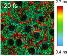
In our research, we aim at visualizing the structure and molecular composition of ocular tissues using both spectral and temporal discrimination. For example, the image shows fluorescence lifetime (FLIM) distribution of retinal pigment epithelium cells of Abca4PV/PV mouse (a model of Stargardt disease in humans). The image reflects differences in the subcellular distribution of endogenous fluorophores in the RPE. Shorter lifetimes (blue-green color) are associated with A2E, a component of lipofuscin. Red granules (longer lifetime) can be associated with retinyl esters.
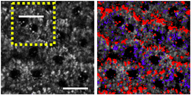
Similar discrimination can be done by looking at the fluorescence spectra. Pixels displaying spectral features of retinosomes were color-coded in red, while those with A2E-like properties were in blue. The analysis shows that retinosomes are predominantly located near RPE cell borders, and some are near cell nuclei. Such studies require specialized ultrafast light sources, scanning ophthalmoscopes, sensitive light detectors, and dedicated software for analysis.
Grazyna Palczewska, Jakub Boguslawski, Patrycjusz Stremplewski, Lukasz Kornaszewski, Jianye Zhang, Zhiqian Dong, Xiao-Xuan Liang, Enrico Gratton, Alfred Vogel, Maciej Wojtkowski, Krzysztof Palczewski, “Noninvasive two-photon optical biopsy of retinal fluorophores”, Proceedings of the National Academy of Sciences 117(36), 22532-22543 (2020).
DOI: doi.org/10.1073/pnas.2007527117
Grazyna Palczewska, Patrycjusz Stremplewski, Susie Suh, Nathan Alexander, David Salom, Zhiqian Dong, Daniel Ruminski, Elliot H. Choi, Avery E. Sears, Timothy S. Kern, Maciej Wojtkowski, Krzysztof Palczewski, “Two-photon imaging of the mammalian retina with ultrafast pulsing laser”, Journal of Clinical Investigation Insight 3(17), e121555 (2018).
One of the methods for evoking plasticity in the visual system is repeated stimulation with appropriate visual stimuli. Repeated exposure to sensory stimuli can induce neuronal network changes in the cortical circuits and improve the perception of these stimuli in the primary visual cortex (V1). The aim of our studies was to investigate the effect of repetitive visual training on the magnitude of visual responses in the primary visual cortex and in the superior colliculus (SC), the subcortical structure of the extrageniculate visual pathway in rats. Our study showed that a three-hour, passive visual training with light flashes enhanced visual responses both at the cortical level and in the superior colliculus. The next part of our study focused on distinguishing which input projection is responsible for the observed training effect in the SC, especially whether the increase of collicular response depends on the enhancement in the V1. The SC receives information both from the retina and from layer 5 of the V1. The experiment with pharmacological blocking of V1 did not suppress training-related plasticity in the SC. These results for the first time identified the superior colliculus as a possible target for training strategies to improve the efficiency of the visual process; e.g., in the case of primary visual cortex injuries.
Author:
dr Katarzyna Kordecka, e-mail: kkordecka@ichf.edu.pl
Publication
Cortical Inactivation Does Not Block Response Enhancement in the Superior Colliculus
Katarzyna Kordecka, Andrzej T. Foik, Agnieszka Wierzbicka and Wioletta J. Waleszczyk
We use cookies to optimize our website and our service.
Functional
Always active
The technical storage or access is strictly necessary for the legitimate purpose of enabling the use of a specific service explicitly requested by the subscriber or user, or for the sole purpose of carrying out the transmission of a communication over an electronic communications network.
Preferences
The technical storage or access is necessary for the legitimate purpose of storing preferences that are not requested by the subscriber or user.
Statistics
The technical storage or access that is used exclusively for statistical purposes.The technical storage or access that is used exclusively for anonymous statistical purposes. Without a subpoena, voluntary compliance on the part of your Internet Service Provider, or additional records from a third party, information stored or retrieved for this purpose alone cannot usually be used to identify you.
Marketing
The technical storage or access is required to create user profiles to send advertising, or to track the user on a website or across several websites for similar marketing purposes.