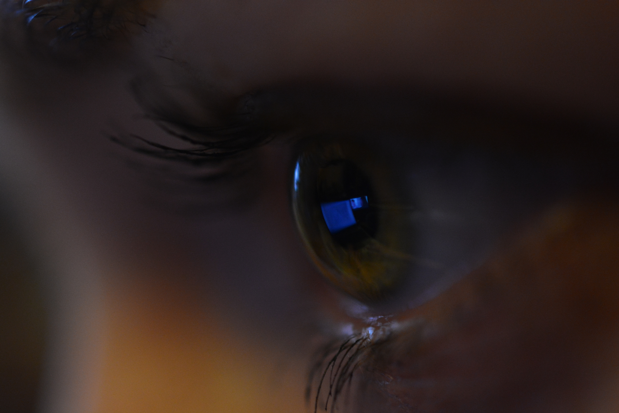
February 4th is World Cancer Day. It gives an opportunity to improve public awareness of this serious and difficult-to-treat disease affecting millions of people around the world.
Cancer is the general name for a group of malignant neoplasms originating from epithelial tissue, although it is commonly used to refer to all malignant tumors that develop in different parts of the body.
As a scientific unit, ICTER (International Centre for Translational Eye Research) has a special interest in malignant neoplasms that develop within the ocular organ. The most common include melanoma, lymphoma, squamous cell carcinoma, and retinoblastoma which affects children.
Melanoma of the eye, as well as the more common melanoma that develops on the skin, originate from melanocytes (pigment cells) which divide rapidly in an uncontrolled manner-. Ocular melanomas are located in various areas of the eyeball and also on the eyelids.
Predisposing factors include light color of the eyes and skin, presence of irregularly shaped nevi on the skin, abuse of sunbathing (including tanning beds), and age (over 50). Research on genetic determinants of uveal melanoma (UM) – one of the eye melanomas performed by many teams identified a number of genes which lead to the development of the tumor when mutated. Among them are: GNAQ, GNA11, PLCB4,CYSLTR2, BAP1, SF3B1, EIF1AX and recently identified MBD4, which encode proteins involved in many biological processes. Eye melanoma diagnosed at early stages of growth can be successfully treated. In contrast, metastasis from the primary tumor to other parts of the body, which is observed in about half of patients, leads to death in 20-30% of cases within 5 years and in as many as 45% within 15 years of diagnosis. Metastasis is the greatest challenge in the treatment of cancers not only of the organ of vision.
To treat melanoma, doctors use radiation therapy, laser treatments and surgery to remove the cancer while preserving the eye. There are also reported cases giving hope for success in the treatment of UM with PD1 inhibitors – compounds used in a promising and relatively new therapy targeting programmed cell death control point. In addition to this therapy, other solutions are being tested in various stages of clinical trials. Apart from immunotherapy, potential drugs are being tested for so-called targeted therapy (targeting e.g. a specific enzyme acting in a cell), epigenetic therapy (targeting DNA modifying proteins), or liver-specific treatment, since it is the main organ where eye melanoma metastases develop. The search for effective drugs continues unabated, many studies have failed, many are still hopeful and others are in the early stages and recruiting volunteers is ongoing.
The same is true for the less common intraocular lymphoma of the eye. Primary intraocular lymphoma is a “variant” of central nervous system lymphoma, whereas secondary ocular lymphoma develops outside the nervous system and occupies the eye through metastasis. The etiology of primary lymphoma is still mysterious. The development of the neoplasm inside the eye is thought to follow the “luring” of appropriate cells circulating in the choroid to the retinal pigment epithelium by chemoattractant such as B-lymphocyte chemoattractant (BLC) or stromal cell-derived factor-1 (SDF-1) present in the choroid. Most primary lymphomas of the eye arise from B-lymphocytes and only a few arise from T-lymphocytes. According to another theory – proposed for patients with compromised immune systems, e.g., AIDS – infectious agents such as Epstein-Barr virus (EPV) cause uncontrolled proliferation of B lymphocytes in the absence of suppressor T lymphocytes.
Many methods are used in imaging lymphoma, or retinoblastoma (described below), including the optical coherence tomography (OCT) – a method developed by the founder of ICTER.
The treatment of lymphoma includes both local solutions – injection of the eye with drugs (Methotrexate, Rituximab), radiation therapy or vitrectomy (surgery performed in the back of the eyeball, on the vitreous body and retina) and systemic solutions. Currently, single-drug chemotherapy and a combination version, where the patient receives mix of different drugs, are used. It is becoming common to combine chemotherapy with radiation. Despite the fact that several medications are approved in the clinic, new ones – more effective and safer – are still being developed.
Another example of eye cancer is retinoblastoma. Patients are young children under the age of 5 who develop cancer in the retina of one or both eyes. Early diagnosis and proper treatment give a chance for full recovery. It is estimated that 40% of cases are caused by genetic factors – inherited or acquired due to somatic mutations. Among genes, mutated in retinoblastoma patients are: RB1, BCOR, MDM4, KIF14, MYCN, DEK, E2F3, CDH11 or RBL2, which encode proteins involved in various biological processes. Moreover, changes in methylation of some genes (e.g. SYK) and dysregulated levels of certain micro RNAs (miR-17~92 and miR-106b~25 cluster) are the cause of retinoblastoma development. The treatment of retinoblastoma depends on the size of the tumor. For small sizes, cryotherapy or laser photocoagulation or thermotherapy is used. Larger tumors are treated with brachytherapy, radiation therapy and chemotherapy – systemic or administered directly to the eye. In extreme cases, when the tumor reaches a significant size, surgery (by removing the entire eye) is required. Untreated retinoblastoma can grow and metastasize to lymph nodes, bone, bone marrow, and the central nervous system. In addition, in the case of genetically affected patients – with mutations in the RB1 gene, there is an increased risk of developing other primary tumors. Such individuals may develop bone, lung, bladder cancer or melanoma, later in life.
Squamous cell carcinoma that develops in various parts of the human eye – usually on the surface of the eyeball – is less invasive than the cancers discussed above and usually does not metastasize to other organs. If left untreated, it can spread within the eye and lead to its loss.
In the fight against eye tumors, it is important to detect them quickly and apply appropriate treatment which, thanks to the achievements of science, are still being improved. For ICTER scientists, this is one of the most important goals.
Author: Magdalena Banach-Orłowska, PhD
Sources:
https://www.zwrotnikraka.pl/swiatowy-dzien-walki-z-rakiem/
https://www.nhs.uk/conditions/eye-cancer/
https://www.cancerresearchuk.org/about-cancer/eye-cancer/treatment/decisions
https://academic.oup.com/jnci/article/113/1/80/5814932?login=false
https://www.nature.com/articles/s41467-018-04322-5
https://www.ncbi.nlm.nih.gov/pmc/articles/PMC5824910/
https://www.ncbi.nlm.nih.gov/books/NBK576390/#article-140272.s2
https://www.ncbi.nlm.nih.gov/pmc/articles/PMC7774148/
https://www.ncbi.nlm.nih.gov/pmc/articles/PMC6034991/
https://www.nhs.uk/conditions/retinoblastoma/
https://www.ncbi.nlm.nih.gov/pmc/articles/PMC7774148/


