

On September 23, 2022, Prof. Marco Ruggeri of the Bascom Palmer Eye Institute visited our centre. His area of expertise includes instrumentation and quantitative imaging technologies for diagnostic and surgical applications in ophthalmology. Having a signed letter of intent with the Bascom Palmer Eye Institute, we discussed potential cooperation looking for joint projects to pursue, especially in the field of ophthalmic procedures. Our scientists Dr. Andrea Curatolo, Dr. Karol Karnowski, Dr. Slawomir Tomczewski, and Marcin Marzejon, M.D., gave Prof. Ruggeri a tour of the laboratories and discussed current research. Prof. Wojtkowski also met with the guest to talk about future projects. During the visit, Prof. Ruggeri gave an interview to our Communications and PR department about the popularization and dissemination of science in the United States and his approaches to promoting research and reaching the widest public with expert knowledge in the field of eye health and new ophthalmic technologies.
Interview with prof. Marco Ruggeri
Please tell us how your specialization translates into improvements in the state of expertise and excellence in vision research.
I work within several niches. First, we want to improve vision in old age so that people can preserve their vision quality later in their life. We first seek to understand why we lose our ability to focus on things up close with age by a condition known as presbyopia. To do so, we are studying the mechanics of accommodation, which is the autofocusing system of the human eye. This is the key part of the process as if we do not know how it works, we will not be able to fix it. We need to find out why we lose this ability as we age so we can counteract it. Since my specialty is optics and imaging, the way I do this is by visualizing and analyzing with our imaging technology what happens inside the eye in real life when we look at near objects and how that changes as we age. We also use this technology to assess the efficacy of the existing procedure to correct this condition, which is important as it provides feedback to manufacturers so that they can improve their products.
I also work on imaging technology for the early detection of eye diseases such as for example, keratoconus. This is important because, with our technology, clinicians will be able to act early and manage the condition in time to maximally preserve vision in patients. But there is more to it because these tools that we develop also provide clinicians with a way to understand whether the current therapies that they are using are effective or not, therefore improving the management of the disease.
As investigators working in translational research, our goal is to move basic science discoveries and technologies more quickly and efficiently into practice. Our vision research center is the ideal place to do so because we are literally located across the street from the hospital of Bascom Palmer Eye Institute, which is one of the largest in the nation. Our approach is to talk to clinicians and identify what the real clinical needs are, and then find a solution. We ask them what scientific discoveries would be game-changers in the field of ophthalmology and would make their life easier, and their feedback is worth focusing on. For example, our institute holds clinical grand rounds every Thursday morning where ophthalmologists confer on complex clinical cases that they discuss by listing different approaches to a given disease or injury. This is one of the best ways to understand what the clinical needs are. You just go there, listen, look at what they are doing, keep quiet, take notes, get ideas and talk to them. I have been doing this for years, and by now, I know most of the ophthalmologists at my hospital quite well. Some of these clinicians eventually became friends. I text them when I need their feedback on a research project, and they text me when they have a new clinical need. I realize this may not be a conventional way of setting scientific priorities, but for me, it proved to be extremely effective. And it has an additional benefit as it is an excellent way to disseminate my scientific work. I also send ophthalmologists my publications, presentations of my scientific work, and share with them the knowledge I explore, primarily driven by a grassroots clinical need.
To recap the life cycle of my work, I first look at a clinical need, and when I identify a meaningful project, I apply for funding to implement it. This is done by preparing a grant application together with a clinician. From the application submission to the funding of large multimillion-dollar from federal entities such as the National Institute of Health takes years, so it is important to be disciplined and act early. Once we receive the funding, I conduct joint research with the ophthalmologists, and the pathway is usually the same; we develop instrumentation and methods, we go into clinical studies on patients and see how it can affect clinical practice. The ultimate goal is to benefit the eye care of patients, so when we reach the end of a research project, and the technology is developed, we start approaching companies to see if they are willing to commercialize our technology bringing it to fruition for patients.
How did your adventure with optical imaging begin, and why did you choose this particular field?
It first started with the eye, even before optical imaging. The eye is a very fascinating part of the body from many points of view. It encompasses mechanical and optical functions, it converts light into electrical signals that travel to the brain and can be used as a window to the rest of the body. I got involved in eye research in Italy during my master’s degree thesis project in electrical engineering – an optical sensor to monitor the glucose concentration in the eye as a potential means of assessing blood glucose. Instead of detecting glucose concentration in the blood, the goal was to measure it non-invasively through the anterior chamber of the eye using an optical technique named polarimetry. That is how I got interested in eye research, but at that time, it was not imaging yet. After graduating, I looked for opportunities to work abroad in the field of measurement technologies applied to eye research. I then found a position as a research associate in the team at Bascom Palmer Eye Institute, developing one of the first implementations of high-resolution OCT retinal imaging for studying the human retina and the retina of small animal models of retinal diseases. It was during that time that I became familiar with the pioneering work of Prof. Wojtkowski on spectral domain OCT imaging. This year marks my seventeenth year at Bascom Palmer Eye Institute.
Are patients in the US aware that more accurate eye imaging methods lead to more effective therapies for eye diseases?
In my experience, not enough.
How do you disseminate your research results and publications?
I participated in the National Alliance for Eye and Vision Research, an organization that promotes advocacy and public education for the eye and vision research sponsored by the National Institute of Health and other federal agencies in the US. Every year they select a few researchers in the field of vision and train them to educate Congressional legislators, the media, and consumers about the value of eye and vision research. For example, we met with government policymakers and explained the importance of allocating taxpayers’ money to eye research and persuading them to promote more research funding for eyesight in the next bill. In the long run, this will save taxpayers money because the funded research will be spent to improve health care.
OCT imaging is a prime example of how technology can lead to significant savings of public funds, with an estimated of more than $10 billion in spending reductions over the last 15 years. The cost saving are the results of clinicians being able to provide more personalized eye care by using OCT to decide when prescription injection are needed to treat some forms of macular degeneration. Thanks to OCT, this process has been optimized by reducing the number of injections needed as well as complication and discomfort to patients.
As for the general population, there are not many channels to disseminate our research and stress its importance, but when it comes to popularizing science, I try to use the same simple language and message as for policy makers, showing the benefits of research applied in ophthalmology. Working in a hospital, I have the great opportunity to explain this directly to patients when they take part to our clinical studies. Other channels to reach the wider public are social media, such as Instagram, LinkedIn, Facebook.
What are the activities of the Bascom Palmer Eye Institute towards the promotion and PR of eye research and science?
Our communications and marketing department regularly publishes a magazine named “Images“ which focuses on the medical and scientific advances at our institution. For example, you can read how our doctors and scientists lead the fight against macular degeneration and how we help babies to see. We have also established a program with the local museum of science in Miami where scientists and clinicians from our institution organize evening seminars to educate the public to our research. Besides that, Bascom Palmer has official channels on social media too, and we are encouraged to work with them to promote our work directly on our institution profiles.
In your opinion, what is the best formula for bringing the nature, importance, and essence of the work of a scientist engaged in eye research to a wider public?
In general, scientists are more comfortable with the conventional and formal ways to disseminate research, such as publications in scientific journals, seminars, and presentations at conferences. While this is important to passing on the benefits of our research to other researchers and professional practitioners, it has a limited outreach for the wider community. The newer generation of scientists is generally doing a better job at promoting the importance of their research on informal channels like social media platforms. Having a marketing department is a great tool for informing the public about research findings. As I explained before, direct contact with patients is helpful. Visits to schools are also a good way to introduce young people to science and get them used to the importance of scientific research. Popular science articles can also be published in the mainstream press, or events can be organized with the local museums for this purpose.
Do you notice any differences in the American and European approaches to science PR, and if so, what are they?
Europeans have made a great effort to promote their research; for example, we observe that scientists are encouraged to have their own laboratory websites or social media accounts. In the US, the promotion of scientists’ work is usually handled by the university’s communications departments. Europe also has great promotional mechanisms in place; for example, when receiving a grant, you are encouraged to advertise your research on a Twitter account. In the US, a special marketing department works for you; they are always looking for news, but we are not pressured, and it is only up to us how much we use their resources to make ourselves known to a wider audience.
What is your greatest professional goal in serving the public?
Generating solutions to improve eye care. The overriding sense of my work is to bring improvements to patients’ vision, ideally moving from research to commercial technology. My dream is that one day people in need can benefit from the technology I have developed.
What are your impressions of Poland and cooperation with Polish scientists so far?
I visited Poland in September of this year for the first time. My impression is that the Polish government is greatly investing significant amounts of resources and money in research. I see that scientific units have access to many grants and other sources of research funding. The cutting-edge technology developed by your centre and other institutions suggests that the level of education is very advanced in your country. Taking part in various conferences where I met Polish scientists, I can confirm that they never failed to present top-notch research. In addition, you are very open and value cooperation. I strongly believe in collaboration among scientists, and I consider that global research should evolve in the direction of international and interdisciplinary cooperation to unite forces and become complementary in what we do. This is the power of today’s science, enabled by modern technology and communication tools.
Thank you very much for the interview and for your visit to ICTER, Professor Marco Ruggeri. We look forward to working with you and can’t wait to start joint scientific projects.
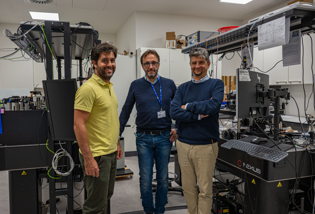
Photo: Dr. Karol Karnowski.
The interview was conducted by the Communication and PR Manager, Dr. Anna Przybyło-Józefowicz.


