05.12.2024

How do we use STOC-T to assess ocular microcirculation? – new paper in Neurophotonics by 3 ICTER research groups

Like a complex network of highways, the retinal microcirculation is a hidden system that powers the life of the eye – delivering oxygen, nourishing tissues, and allowing cells to function without disruption. New research conducted by ICTER (International Centre for Translational Eye Research) scientists will enable us to track every “movement” on these microscopic roads using the STOC-T (Spatio-Temporal Optical Coherence Tomography) technique. This offers the opportunity to understand the mechanisms of retinal function and discover how microcirculation disorders herald the onset of neurological and ophthalmological diseases.

Retinal microcirculation and hemodynamics provide valuable information on neurovascular diseases, as many diseases of the central nervous system (CNS) can manifest themselves through changes in the retina. Given that about 80% of external information is processed through visual perception, understanding the structure and function of the retina, vascular hemodynamics, and neurovascular coupling (NVC) is of paramount importance.

Now, ICTER scientists have used spatio-temporal optical coherence tomography (STOC-T) to assess retinal microcirculation. It turns out that the STOC-T technique, which uses fast near-infrared tomographic imaging, offers the possibility of visualizing even the smallest capillaries in real-time. Unlike other techniques, such as ocular angiography (angio-OCT) or Doppler tomography, STOC-T allows for obtaining 3D images of the entire structure of the retina and choroid with high temporal precision. Additionally, the use of digital aberration correction and a specially designed optical system allows for obtaining images unaffected by refractive errors, which is particularly important for imaging small structures, such as the mouse retina. The results were published in the journal Neurophotonics in a paper entitled “In vivo volumetric analysis of retinal vascular hemodynamics in mice with spatiotemporal optical coherence tomography.”

What connects STOC-T and ocular microcirculation?

Spatio-temporal optical coherence tomography (STOC-T) is an advanced optical tomography method that allows for obtaining three-dimensional images of tissue microstructures in real time with high temporal resolution. In turn, ocular microcirculation is, broadly speaking, a network of small blood vessels supplying the retina and choroid, allowing for the proper functioning of photoreceptors. Adequate blood flow is essential for the delivery of oxygen and nutrients and the removal of metabolic products.

Retinal microcirculation research is becoming particularly important in the context of the increasing number of neurodegenerative and ophthalmological diseases. Disorders such as Alzheimer’s disease, Parkinson’s disease, and multiple sclerosis, as well as eye diseases such as glaucoma or diabetic retinopathy, are often associated with microcirculation disorders that can be visible at the retinal level even before neurological symptoms appear. The use of STOC-T allows precise monitoring of hemodynamic changes in the retina, which can help in the early detection of pathologies and the development of new therapeutic methods.Retinal blood flow can be quantitatively monitored in vivo using laser speckle flowgraphy (LSFG), a technique that generates angiographic contrast from speckle variance, enabling full-field (arbitrary units) blood flow measurements with high temporal resolution. Alternatively, laser Doppler flowmetry (LDF) can measure blood flow and mean velocity in relative units, and its extension, laser Doppler holography (LDH), can estimate pulsatile retinal flow in the lateral field of view (FOV) with millisecond resolution. These techniques cannot perform deep slices, making the influence of choroidal flow unclear. It would be beneficial to analyze choroidal hemodynamics separately from the internal retinal hemodynamics and with high temporal resolution, which is what STOC-T allows.

Groundbreaking observations and a chance for new therapeutic options

The study aimed to implement the STOC-T technique, previously developed by ICTER scientists, for monitoring retinal microcirculation and neurovascular coupling (NVC). Now, it was possible to obtain detailed images of different layers of the mouse retina, such as the neurofibrous layer (NFL), the inner plexiform layer (IPL), the inner and outer photoreceptor segments (IS/OS) and the choroid. These images allow for the observation of both larger blood vessels on the surface and the more complex network of capillaries in deeper layers, such as the IPL.

Analysis of the STOC-T signal amplitude allowed for the differentiation of arterial and venous pulsations in the mouse retina. In particular it was found that the pulsation in the venous vessels is delayed by an average of 29 milliseconds about the arteries, which allows for the identification of phase differences between these vessels. This pulsation time delay between arteries and veins is crucial for understanding the different roles these vessels play in microcirculation. STOC-T allows tracking of tissue displacements induced by the pulse wave as it travels through the retinal layers. These micromovements are measured in nanometers and observed mainly around arteries and veins, with modulation amplitudes ranging from 100 to 150 nanometers.

Measurement of the blood pulse wave velocity (0.35 mm/s) in the capillaries of the outer plexiform layer (OPL) and tissue displacements induced by vessel pulsation (up to 150 nm) provided data on the biomechanical properties of the different retinal layers. This analysis revealed differences in the biomechanical response to pulsation between layers, which is particularly valuable for NVC studies. Although the system is limited in recording pulse wave velocity in larger vessels due to the field of view and pulse wavelength, it remains highly effective in analyzing blood flow in capillaries.

Mapping of tissue shifts in time caused by vascular pulsation revealed that retinal layers exhibit periodic expansion and contraction synchronized with vascular pulsation. These observations, with an amplitude of 100-150 nm, provide important information on tissue elasticity and biomechanical properties of the retina, enabling further studies on neurodegenerative diseases in which the preservation of microcirculation and vascular elasticity may be impaired.

New quality in imaging of ocular hemodynamics

Studies conducted using the STOC-T technique provided detailed data on the hemodynamics and biomechanics of the mouse retina, which opens new diagnostic and therapeutic perspectives. The possibility of noninvasive monitoring of retinal blood flow and precise analysis of phase differences between venous and arterial pulsations may be crucial in the detection and treatment of many neurological and ophthalmological diseases. Ocular microcirculation, being a hidden highway supplying the retina with oxygen and nutrients, is crucial not only for the health of the eyes but also for the condition of the entire nervous system.

This publication is the result of the fruitful cooperation of three ICTER groups: POB, IDoc, and OBi, which emphasizes its interdisciplinary nature. These teams, combining their unique experiences and expertise, created the foundations for innovative solutions described in the publication. Such a combination of knowledge from different research areas is a key element in the search for innovative answers to contemporary challenges, which is one of the pillars of ICTER.

Authors of the paper “In vivo volumetric analysis of retinal vascular hemodynamics in mice with spatio-temporal optical coherence tomography”: Piotr Węgrzyn, Wiktor Kulesza, Maciej Wielgo, Sławomir Tomczewski, Anna Galińska, Bartłomiej Bałamut, Katarzyna Kordecka, Onur Cetinkaya, Andrzej Foik, Robert J. Zawadzki, Dawid Borycki, Maciej Wojtkowski, Andrea Curatolo.

Press note author: Scientific Editor Marcin Powęska.

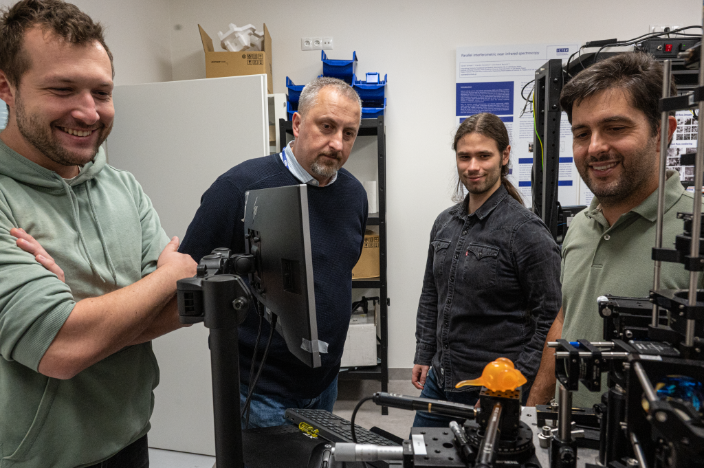
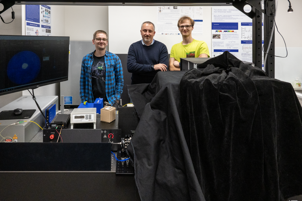
The pictures portray the first author of the paper: Piotr Węgrzyn. Photos by Dr. Karol Karnowski.

23.10.2024

Key to the invisible world found. ICTER scientists decipher two-photon vision

Two-photon vision is a novel method with great potential for the future of ophthalmic diagnostics. Although it has many advantages, it requires improvement in key areas. ICTER scientists have taken a step forward by improving this technology and opening up new perspectives in ocular medicine.

Imagine that instead of viewing an image through a lens, you look through a kaleidoscope that focuses invisible light to obtain a new range of colors. The photon, this ephemeral messenger of light, usually appears alone, but here it appears in a duet, which is the basis of two-photon vision. This extraordinary phenomenon in which the human eye, instead of perceiving traditional light, receives pulses of infrared lasers, the gateway to the invisible world.

However, the key to them is measuring the brightness of two-photon stimuli, which until now was only possible for visible light. ICTER scientists have made a breakthrough and determined the luminance value for infrared using photometric units (cd/m2). Thanks to this approach, it was possible to link the luminance of two-photon stimuli to a new physical quantity related to perceived brightness: the two-photon retinal illumination.

Research conducted by scientists from the International Centre for Eye Research (ICTER) with the participation of PhD student Oliwia Kaczkoś, PhD Eng. Katarzyna Komar and Prof. Maciej Wojtkowski have shown that the luminance of a two-photon stimulus can reach almost 670 cd/m2 in the safe range of laser power for the eye. The result is the paper entitled “Method for the determination of the luminance of two-photon vision stimuli,” published in the journal Biomedical Optics Express.

From left: Oliwia Kaczkoś and Dr. Katarzyna Komar. Photo by Dr. Karol Karnowski.

Seeing the Invisible World

The human eye can receive stimuli from the surrounding world in the form of electromagnetic waves in the range of about 380 nm to 780 nm (from violet to red). Waves outside this range, such as infrared (above 780 nm) and ultraviolet (below 380 nm), are invisible to us without special devices, although they can affect the senses in other ways.

Every vision process follows the same path when a photon of visible light is absorbed by the visual pigment of the photoreceptor in the retina (the light-sensitive part of the eye). This event initiates a series of chemical reactions, as a result of which a quantum of light is converted into an electrical signal, processed in the brain.

Two-photon vision is a phenomenon in which the human eye can perceive ultrashort pulses of infrared lasers with a wavelength in the range of 800-1300 nm by absorbing two photons. This process causes isomerization of visual pigments, which leads to the perception of light with a wavelength corresponding to half the infrared wavelength. Although these lasers are outside the visible range of the spectrum, their effect on visual pigments allows infrared light to be recorded as different colors.

Two-photon vision differs from single-photon vision primarily in the way light is absorbed. In single-photon vision, each photon with a specific energy is absorbed by molecules in the eye, which allows light to be perceived in the visible range. In two-photon vision, on the other hand, two photons with half the energy are simultaneously absorbed by visual pigments, which leads to the perception of light with half the wavelength, which theoretically should not be visible.

Furthermore, the brightness of the two-photon stimulus varies with the square of the power of optical radiation, so light scattered in the eye will not be perceived. Brightness also depends on the focus of the beam on the observer’s retina – the received stimuli are sharper and have better contrast than in the case of “normal,” single-photon vision.

ICTER scientists have long been studying the phenomenon of two-photon vision, were the first in the world to describe it, and now they have made another groundbreaking discovery.

On the picture: Oliwia Kaczkoś. Photo by Dr. Karol Karnowski.

A novel method for determining the brightness of two-photon stimuli

Two-photon vision shows potential in two key areas: medical diagnostics and virtual/augmented reality (VR/AR). It can be used for advanced diagnostic tests, especially in neurology and ophthalmology, where infrared pulses allow for safe monitoring of visual functions without the need to use visible light. On the other hand, this phenomenon allows the creation of new, realistic visual experiences by manipulating light stimuli from the infrared range, opening up new possibilities in interaction with virtual images (VR/AR).

All future applications of this phenomenon require knowledge of the luminance of two-photon stimuli, but the luminous efficiency function V(λ) outside the visible range is unknown. A non-standard approach to quantifying the luminance of two-photon stimuli is necessary, e.g., using infrared – which is what ICTER scientists did.

The method presented in the paper allowed the expression of the brightness of two-photon stimuli in photometric units. Thanks to the measurements performed, the scientists were able to demonstrate the relationship between the power of the infrared beam and the power of the visible beam, which was subjectively adjusted so that both were perceived as having the same luminance. Using the relationship between the power density of the VIS laser and the luminance of the projected stimuli, it was possible to determine the subjective luminance of the infrared stimuli using photometric units (cd/m2). These results emphasize the nonlinear nature of two-photon vision, which is in agreement with previous studies.

“The study aimed to develop a repeatable method for determining the brightness of stimuli for two-photon vision. Standard methods do not allow this to be done outside the visible spectrum of light, but our research opens the door to achieving this goal, which is necessary for further research and development of applications of this phenomenon in medical diagnostics and augmented reality (AR) and virtual reality (VR) technologies. The new approach will also enable comparison of the brightness of two-photon stimuli with traditional displays based on standard, single-photon vision”. – says Oliwia Kaczkoś, ICTER PhD student and optometrist, lead author of the study.

From left: Dr. Katarzyna Komar and Oliwia Kaczkoś. Photo by Dr. Karol Karnowski.

A platform for further discoveries

The result of the research is the proposal of a completely new physical quantity, called two-photon retinal illumination, which is sufficient to describe systems emitting two-photon stimuli. This relationship allowed the prediction of the luminance values ​​of two-photon stimuli, which could reach 670 cd/m2 in the safe laser power range of the human eye without adaptive optics (AO) correction.

Moreover, ICTER scientists documented twice the repeatability for measurements made on a background with a luminance of 10 cd/m2. This is crucial for the development of future technologies, such as two-photon retinal displays, which could be used in augmented reality (AR) glasses or in advanced diagnostic tools such as two-photon microperimetry.

Authors of the paper “Method for determination of luminance of two-photon vision stimuli”: Oliwia Kaczkoś, Agnieszka Zielińska, Jacek Pniewski, Maciej Wojtkowski, and Katarzyna Komar.

Author of the press release: Marcin Powęska.

Photos: Dr. Karol Karnowski.


ICTER, or the International Centre for Translational Eye Research, is a research, development and innovation centre at the Institute of Physical Chemistry of the Polish Academy of Sciences (IChF), located in Warsaw, Poland. ICTER was established in 2019 to develop cutting-edge technologies to support the diagnosis and therapy of eye diseases, based on funding from the International Research Agendas Programme of the Foundation for Polish Science, co-financed by the European Union – European Regional Development Fund. The centre is currently implementing the MAB FENG grant of the Foundation for Polish Science. In 2024, IChF has won a prestigious grant under the Teaming for Excellence / WIDERA program of Horizon Europe, which will enable ICTER unit to develop into a European centre of excellence. The centre’s website is: www.icter.pl.

02.10.2024

The technology that conquered rapid eye movements

A special camera, laser, and computer will provide an even more precise view of what is happening inside our eyes, according to researchers from the ICTER research centre. The new examination is painless and takes only 8.6 milliseconds, which is equivalent to the time it takes for a bee to flap its wings twice. This technology aims to enhance the detection of eye diseases at their early stages.

The technology, known as STOC-T, developed by scientists, has the potential to replace today’s OCT examination, commonly referred to as eye tomography. The team behind this new technology is led by Professor Maciej Wojtkowski, the head of ICTER. Notably, a quarter of a century ago, he created the first prototype of the OCT device, which is now used in ophthalmology clinics worldwide.

Capturing the eye in stillness

“In eye examinations, time is crucial. The eye constantly makes small, unconscious movements. Even though the previous technology only required a few seconds for an examination, each of these movements introduced noise, reducing the clarity of the results,” explains Professor Maciej Wojtkowski.

The development of digital technologies provided a solution. Researchers utilized a camera capable of capturing up to 60,000 frames per second, which is over a thousand times faster than standard smartphone cameras. As a result, the examination time was reduced to less than 0.01 seconds.

“During this imperceptible fraction of a second, our system illuminates the eye with multiple laser light waves. The camera captures hundreds of frames showing how the light propagates within the cells of the eye. These frames are then superimposed and analyzed by our software, which generates a precise image of the eye’s layers,” says Professor Maciej Wojtkowski.

This approach provides ophthalmologists with a detailed image of the retina, enabling them to detect early signs of various eye diseases, including glaucoma, macular degeneration, and diabetic retinopathy. The increased precision of diagnosis is particularly important in Poland, where one ophthalmologist serves approximately 10,000 patients. According to the latest report from the National Health Fund, more than half a million Poles are currently on waiting lists for ophthalmology appointments. Early diagnosis allows for quicker treatment, which, in many cases, can prevent vision loss.

International support

The ICTER research centre is part of the Institute of Physical Chemistry of the Polish Academy of Sciences. Its scientists combine knowledge from physics, biology, chemistry, engineering, and medicine. This interdisciplinary approach allows them to create new methods for examining and treating vision.

The developed technology is one of many steps planned by researchers. Their primary goal is to find a technological solution that will facilitate faster access to specialized ophthalmic care. Their efforts have been made possible by a prestigious grant awarded under the “Teaming for Excellence” program in Horizon Europe. They have secured €30 million in funding, half of which comes from the European Commission, with the remaining amount provided by the Foundation for Polish Science and the Polish Ministry of Education and Science.

With this funding, ICTER will establish a Centre of Scientific Excellence in Warsaw — a place where science meets practice, and research is transformed into concrete medical solutions. Partners in this project include the Institute of Ophthalmology at University College London and the Institut de la Vision at Sorbonne Université.

02.04.2024

Stargardt’s disease has many names. Different varieties require different approaches – research from ICTER published in PNAS Journal

Stargardt’s disease is a rare eye condition that affects both children and adults, in many cases leading to blindness. It has several different varieties, and new research indicates that each requires a slightly different therapeutic approach. This key discovery may contribute to the development of effective methods to combat this currently incurable disease.

Initially, patients experience a sudden loss of sharp and central vision. The disorders progress, often making normal functioning difficult, including reading or face recognition. For many people, just looking at a light source is painful. After some time, the disease may stabilize, but often the damage to the macula is so significant that vision loss occurs.

The above description concerns Stargardt’s disease, a disorder that affects approximately 1 in 10,000 people, most often children between 8 and 12 years of age. There is no cure for this macular dystrophy, but understanding the mechanisms that occur in photoreceptors during the disease is crucial for potential therapeutic interventions (several drugs are being tested). It turns out that different types of Stargardt’s disease – although they have a similar cause – respond differently to different classes of drugs.

Research conducted with the participation of scientists from the International Centre for Translational Eye Research (ICTER) at the Institute of Physical Chemistry, Polish Academy of Sciences, including Dr. Marcin Tabaka, Dr. Andrzej Foik, Dr. Damian Panas, Dr. Jagoda Płaczkiewicz, and Dr. Katarzyna Kordecka, in cooperation with the Center for Translational Vision Research at the Gavin Herbert Eye Institute (CTVR) from UC Irvine in California under the supervision of Prof. Krzysztof Palczewski, resulted in a paper published in PNAS entitled “Distinct mouse models of Stargardt disease display differences in pharmacological targeting of ceramides and inflammatory responses”, which increases our understanding of Stargardt’s disease.

Stargardt’s disease is not just one disease

Lipids are an important component of the biological activity and physiology of most cells in the body, and in particular, they play a key role in the functioning of the retina. It has been confirmed that disturbed lipid homeostasis occurs in various degenerative retinal diseases, although still little is known about lipid profiles in normal and diseased retinas.

Stargardt’s disease is characterized by the accumulation of a non-degradable derivative of the visual pigment – lipofuscin – in the retinal pigment epithelium (RPE), which causes its atrophy. From the first description of Stargardt’s disease until recently, its diagnosis was based on the evaluation of the phenotype by eye examination, but since the advent of genetic testing, the picture of the condition has become more complete.

What was originally thought to be one disease is probably at least three variants (STGD1, STGD4, STGD3), each associated with a different genetic change. Therefore, it is extremely difficult to clearly define what Stargardt’s disease is, let alone think about potential treatments.

The most common form of Stargardt disease – STGD1 – is caused by biallelic variants of the Abca4 gene (i.e. autosomal recessive), and their exact genotype (i.e. combinations of both variants) is of great prognostic importance as to the age of disease onset and its progression. The frequency of Abca4 allele carriers in the general population is 5-10%, and different gene combinations influence the age of onset and the severity of the disease itself. Furthermore, disease severity is inversely related to ABCA4 function, and mutations in this gene are thought to play a role in other diseases such as retinitis pigmentosa, rod and cone dystrophies, and Age-Related Macular Degeneration (AMD). Macular Degeneration).

Biochemistry of Stargardt’s disease

Two genes encoding lipid-processing proteins have been associated with inherited retinal degenerative diseases. Mutations in the gene encoding an enzyme called ELOVL4 elongase, involved in building long-chain fatty acids, have been associated with autosomal dominant AMD-like Stargardt disease – STDG3 variant. Patients suffering from this disease at a young age experience loss of central vision. In turn, the disease in people suffering from STGD4 has been associated with mutations in the Prom1 gene. This disease variant was discovered in 1999 and little is known about its cause, except that there is a mutation at the p.R373C site, which causes dystrophy of cones and rods.

However, the most common form of Stargardt disease – STDG1 – is quite well understood. Patients showed a progressive bilateral appearance of yellow-orange spots (lipofuscin) in the macula, where the density of cones is highest. Such changes cause atrophy of the RPE and the death of photoreceptors in this region, resulting in vision loss. Lipofuscin accumulation in the RPE is a characteristic finding in the eyes of STGD1 patients and the mouse model of Stargardt disease.

Previous studies have shown that ABCA4 is expressed in the photoreceptor outer segment disc (POS disc) and the plasma membrane of RPE cells. ABCA4 transports a retinal phospholipid compound known as N-retinylidene-phosphatidylethanolamine (N-ret-PE) upon photoexcitation, allowing its removal from receptor cells – similarly to RPE cells, where N-ret-PE is transferred to lysosomes or phagosomes. In people with Stargardt disease (STDG1), N-ret-PE accumulates in POS discs and leads to the formation of the precursor A2E bisretinoid (A2PE). Inside the RPE lysosomes, A2PE is converted into A2E – a compound that cannot be broken down by any enzymes in the body. A2E is believed to react with other lipids and convert to lipofuscin deposits.

According to the current knowledge, over 100 mutations affect ABCA4 functions and are associated with a wide spectrum of Stargardt disease phenotypes. The suggestion that mutations in different domains of ABCA4 may affect the RPE and photoreceptors differently is tempting, meaning that ABCA4 has slightly different functions in them. This, in turn, opens a window into the potential therapeutic mechanism associated with lipid-lowering drugs.

Therapeutic hopes

Although there is no cure for Stargardt’s disease, work is underway on several therapies aimed at limiting or completely stopping the progression of this disease. The team led by Prof. Palczewski at CTVR and ICTER decided to investigate two alternative mouse models of functional ABCA4 deficiency to model the heterogeneity of mutations in patients with Stargardt disease and to see whether a different therapeutic approach might be necessary to stabilize retinal health in each case.

Using a combination of molecular techniques, the researchers examined Abca4 knockout mice (knock-out) and mice with additional Abca4 (knock-in). The researchers compared the two strains to determine whether they exhibited differential responses to factors that affect cytokine signaling and/or ceramide metabolism, as changes in either of these pathways can exacerbate retinal degenerative phenotypes.

Abca4 knockout and Abca4PV/PV knockout mice showed different responses to a ceramide-lowering drug and an immunomodulatory drug. The two mouse models were found to have divergent levels of baseline cellular stress and signaling, which are exacerbated in the early stages of light-induced retinal degeneration. Most importantly, these side effects can be alleviated prophylactically with the use of an immunomodulatory drug, thus “slowing down” the course of Stargardt’s disease.

“We found varying degrees of response to maraviroc, a known immunomodulatory CCR5 antagonist, and to the ceramide-lowering agent AdipoRon, an agonist of ADIPOR1 and ADIPOR2 receptors. Our phenotypic comparison of two different mouse models with the Abca4 mutation sheds light on potential therapeutic options previously unexplored in the treatment of Stargardt’s disease and provides a substitute test for assessing the effectiveness of gene therapy” – says Dr. Marcin Tabaka, leader of the ICTER Computational Genomics Team.

It is worth mentioning that in an earlier study, also published in PNAS, entitled “Stress resilience-enhancing drugs preserve tissue structure and function in degenerating retina via phosphodiesterase inhibition”, molecules that activate biological mechanisms of stress resistance were examined. This, in turn, may result in the development of a new class of pharmaceuticals, the so-called drugs that increase stress resistance (SRED) with potentially wide clinical significance for combating pathological retinal changes, not only Stargardt’s disease. A major contribution to the research work was made by the ICTER Computational Genomics Team, which analyzed single-cell sequencing data, enabling the identification of universal molecular mechanisms involved in age-related eye diseases and hereditary retinal degenerations.

The latest study makes a case for using transgenic knock-in mouse lines that have mutations analogous to those in humans. It also serves as a precursor to adopting these models to test genome editing methods that can precisely correct genetic mutations. Thanks to these efforts, Stargardt’s disease, which is today considered “incurable”, may become a disease that can be controlled and, as a result, save eyesight.

Author of the press release: Marcin Powęska.
Image: Deposit Photos.

Cited papers:
https://doi.org/10.1073/pnas.2314698120
https://doi.org/10.1073/pnas.2221045120

13.12.2023

“Optoretinography is the future of ophthalmology, and the knowledge from ICTER is utilized by top specialists” – interview with Prof. Robert Zawadzki from UC Davis

Thanks to medical progress, we can cure more and more vision-related diseases and the bottleneck of successful ophthalmological interventions is diagnostics. The stage at which changes in the retina are detected directly translates into the patient’s chances of recovery. One of the most innovative and fastest-growing ophthalmological techniques is optoretinography (ORG), the leader of which in Poland is Prof. Maciej Wojtkowski from ICTER. Many centers around the world research ORG, and many use the treasure trove of knowledge of ICTER scientists.

One of the most important research centers specializing in ORG in the United States is the University of California at Davis (UC Davis), where Prof. Robert Zawadzki – a graduate of the Nicolaus Copernicus University in Toruń and a long-time collaborator of ICTER – has worked for about 20 years. We asked him what he does at UC Davis; how his research can translate into patients’ health; what are his feelings after visiting ICTER and why cooperation between centers from all over the world is crucial for the future of ophthalmology.

Please tell us what project you are working on and with whom during your current visit to ICTER.

Yes, this is my follow-up visit to ICTER at the invitation of Prof. M. Wojtkowski. Our plan during my previous visit was to assist in two research projects. One involved setting up and testing a fundus camera, which was designed for the STOC-T system and is now used for functional eye imaging. This research was conducted in collaboration with Dr. Andrea Curatolo’s team, with Wiktor Kulesza and Piotr Węgrzyn. During that stay, we managed to obtain an image of the mouse eye fundus using the camera, which could be used to determine the precise location of the retina for subsequent functional measurements using STOC-T. The second project involved cooperation with Dr. Michał Dąbrowski, that focused on assisting in the two-photon fluorescence imaging of the retina. Here, I also helped Mr. Michał correct the image from the auxiliary single-photon scanning light ophthalmoscope system, for two-photon measurements. In both the STOC-T and two-photon projects, the primary scientific instruments did not have real-time high-quality images, and this is where assistance was needed to build systems that would help align the eyes during the examination. During my current stay, I also participated in the CRATER conference and then focused mainly on working with Dr. Andrea Curatolo’s team. We collaborated on a manuscript describing the STOC-T measurement system for mouse imaging and its applications. Additionally, we discussed issues related to finding permissible light exposure limits for STOC-T measurements on experimental animals, as well as the details of the STOC-T optical system and the potential impact of various components on the measurement system’s resolution.

This is not the first time you have come to our centre. Please tell us briefly about what has been achieved during your previous visits and in what direction further cooperation with ICTER researchers is going.

Indeed, these visits are a continuation of my previous ones. I have previously collaborated with the same teams. During one of my first visits, taking place over two years ago, Dr. Michał Dąbrowski was still building his system, so our collaboration was limited to assisting in selecting certain optical components, which were needed for the experiments we conducted in 2022. In the case of Dr. Andrea Curatolo, during my first visits, we collaborated on the design, construction, and initial setup of the system. I hope that both projects will continue to develop and, in the case of Dr. Curatolo, allow for functional measurements at the retina in laboratory animals using STOC-T, and in the case of Dr. Dąbrowski, be useful for two-photon fluorescence measurements that may provide us with more information about diseases of photoreceptors and retinal pigment epithelium, cells containing majority of fluorescent molecules in the eye.

From the left: Piotr Węgrzyn, Prof. Robert Zawadzki, Wiktor Kulesza and Dr. Andrea Curatolo at ICTER’s lab.

What areas do you specialize in and what are the unique effects of this non-obvious combination in practice?

I specialize in the field of biomedical engineering or bio-photonics, specifically focusing on building and utilizing systems for functional measurements in the eye, particularly on the retina, in both humans and experimental animals. The outcomes of my work involve the development of new devices that enable the measurement of functional changes at the cellular level resulting from disease-related changes or age-related alterations. In the future, these methods may contribute to better diagnostics and the assessment of the effectiveness of gene or stem cell therapies in such cases.

Please tell us about your research and work at UC Davis.

I have been at UC Davis for about 20 years now, and currently, I’m a professor in the Department of Ophthalmology & Vision Science. I’m a member of two research groups there. One group is focused on testing and building clinical research devices; it’s called CHOIR, which stands for the Centre for Human Ophthalmic Imaging Research. The other research group I’m part of, EyePod Small Animal Ocular Imaging Laboratory, is involved in small animal ocular imaging. We design and create new devices for structural and functional eye measurements, mainly in mice and small experimental animals. The research we conduct aims to develop new methods that can be useful for both clinical doctors and scientists working on fundamental medical research, where new methods such as gene and stem cell therapy are being developed. We are one of the groups that helps other research teams test their innovations more effectively and identify potential issues faster while also aiding them in discovering new directions for the development of these various therapies.

How can you translate your research results into measurable and useful applications for patients?

Our research has the potential to be useful for patients in the following two scenarios. The first is the development of devices to enhance the diagnosis of eye diseases, improving these methods to the extent that even for individuals who do not yet exhibit any objective changes in their vision, it will be possible to determine whether there are any underlying changes. This is particularly crucial among individuals with genetic predispositions that put them at an increased risk. Knowing a person’s specific genetic defect can help tailor diagnostic methods to identify functional changes in certain cells, potentially allowing for prevention or at least a delay in the progression of the disease, given the current state of the medicine. In cases where these therapies are expensive, this is indeed a significant aspect. The second direction of our research is to confirm whether the methods used to treat patients are effective. In this scenario, if we find no changes, the doctor may choose another method that produces better results. This aspect is perhaps more tangible for patients. Of course, our research is also crucial in the implementation of new therapeutic methods as it accelerates the development of these therapies.

Please tell us how the optoretinography technology developed with ICTER is state-of-the-art, where in the world is it currently being developed, and what is your unique contribution to its development?

The technology of optoretinography, referred to as the method of measuring the functional response of the retina to light stimulation, has unique diagnostic potential and is, therefore one of a kind, it is currently being researched in various laboratories to understand how the signals measured using ORG can be linked to known physiological functions of individual retinal neurons. In Europe is primarily being developed by groups like ICTER led by Maciej Wojtkowski, a strong group in Germany under Geron Huettmann, and another group in Paris, lead by Kate Grieve. In United States, we have groups at UC Davis, University of Washington led by Ramkumar Sabesan, Indiana University led by Don Miller. There are also groups at the University of Illinois Chicago, the University of Wisconsin, the University of Pennsylvania and at Stanford University, just to name a few. All these groups focus on various aspects of ORG. My contribution to the development of this method involves e elucidating the physiological factors related to these signals. We have been able to confirm that changes in retinal water content are responsible for a portion of the signals we measure. This is a secondary effect following photoreceptor light activation but is related to water. Additionally, in our group, which develops devices for clinical research, we aim to create models that would enable us to validate our results, making it easier to determine the main characteristics of the optoretinography signal. Our research is directed towards finding better methods for optoretinographic measurements, discovering what truly influences the signal we measure. Note that we mainly measure changes in the thickness of certain retinal layers and alterations in light scattering. We are also developing methods to model these signals and more easily find correlations between the parameters of these curves and various eye diseases.

From the left: Dr. Michał Dąbrowski, Prof. Robert Zawadzki, and Bartłomiej Bałamut at ICTER’s lab.

Please specify how your career and approach to science have been influenced by the different locations and units where you have worked so far: undergraduate and graduate studies at UMK in Toruń, PhD in Vienna, and work at UC Davis.

My undergrad studies at Nicholaus Copernicus University (UMK) in Toruń were indeed essential for me to find myself where I am now, but, as in most cases, one’s career path and life journey are highly individual and challenging to replicate for others. They are often the result of certain coincidences and opportunities that have appeared on my path, some of which I was able to seize while others eluded me. However, my undergraduate studies at UMK were crucial in gaining fundamental knowledge in experimental physics and the use of computers in physics. They laid the foundation for my understanding of the scientific alphabet, so to speak. Then, during my master’s studies, I had the incredible fortune to start collaborating with Prof. Andrzej Kowalczyk, who, in the late 1990s, had a European Tempus grant for sending young students on various internships. In my case, I had the opportunity to intern at a university in Vienna, where I first encountered the method, I currently work on, Optical Coherence Tomography (OCT), and I also met one of its inventors, Prof. Adolf Fercher. After completing my master’s degree, I received an offer to pursue a Ph.D. in Vienna under Prof. Fercher, which is when I embarked on my doctoral studies. This experience allowed me to become proficient in both the OCT method and the field of biophotonics, as well as understand how to design devices for studying the eyes and other organs and how to apply data analysis methods. Therefore, my doctoral work was instrumental in building the knowledge needed for what I do now. After completing my Ph.D. in Vienna, I worked briefly as an assistant at UMK, and then, after about six months, I received a job offer at UC Davis as a Postdoc in John Werner Laboratory. It was there that I engaged in a significant project funded by the National Eye Institute, which involved building the world’s first system that combined adaptive optics with OCT. The knowledge I had gained during my doctoral studies, particularly in using OCT to study corneal shape and detect eye aberrations, proved to be ideal for this project, as I already had a foundation in ocular aberrations and understanding the function of the eye as an imaging element, as well as the basics of OCT. This experience in Vienna had also allowed me to become familiar with then up-and-coming detection technique known as Fourier Domain OCT. So, when I went to UC Davis, I had all the knowledge necessary to complete this project, which involved creating the first working adaptive optics OCT (AO-OCT) system, and we demonstrated the first images with cellular resolution on the retina. Throughout the years working at UC Davis, I maintained collaborations with groups in Toruń, led by Prof. Maciej Wojtkowski, and in Vienna. As these eye imaging methods evolved, we contributed to their development, primarily focusing on optical coherence tomography angiography, a method for non-invasively measuring blood flow in the eye. We also worked on methods that combined several different imaging techniques such as OCT with SLO, which are utilized by many modern systems for retinal eye imaging. About 12 years ago I began using these systems for measurements in experimental animals. This was made possible through collaboration between UC Davis Department of Ophthalmology (Prof. John Werner’s group) and the Department of Physiology, where Prof. Edward Pugh was involved. Through our collaboration with Ed Pugh, we created the EyePod team, which focused on studying the retina in experimental animals. It was around 2015 when we began working on ORG, or optoretinography. Thus, I continue to work in the same field, which I have been engaged in since my master’s studies, namely development and application of OCT in Medicine. I was able to do it by constantly applying the latest research method and technology. I have also been able to continuously expand my knowledge to keep my work as interesting and attractive as possible for these new emerging application fields.

What would you like to pass on to fellow scientists involved in eye research and the development of new ophthalmic therapies?

I would like to say that despite the fact that our new research methods and therapies seem very advanced, there are still many things we don’t know and cannot measure yet. I suspect that there is still a lot of work ahead of us to make these methods we are working on clinically available. Just as all these fields are still evolving, I would recommend young scientists to look at the current issues related to eye research. Perhaps even their individual experiences can be crucial in finding further solutions. So, the development of novel structural and functional assessment of the eye is something worth continuously engaging with.

Thank you very much for this interview, Prof. Robert Zawadzki. We eagerly anticipate further fruitful collaboration in the future.


Special thanks to all the ICTER scientists who participated in the photo session at our laboratories.

The interview was conducted by Dr. Anna Przybyło-Józefowicz (September 2023)

Title, introduction, and social media material: Journalist Marcin Powęska

Pictures: Dr. Karol Karnowski

15.09.2023

ICTER’s Overview Report 2019-2023

We are pleased to present the ICTER’s Overview Report for the years 2019-2023.

This report offers an insightful look into our organization:

  • Discover our dedicated team and their collaborative spirit.
  • Explore our unwavering mission to advance global eye health.
  • Learn about the funding that fuels our initiatives.
  • Gain insights into our impactful grant projects.
  • Trace our journey through a brief overview of our history.
  • Delve into our contributions to the field through publications.
  • Understand our communication and outreach strategies.
  • Explore our fruitful collaborations with industry.
  • Connect with our network and ecosystem.
  • Meet our diverse research groups and their focus areas.
  • Stay informed about our hosted events and notable visitors.
  • Join us in celebrating the recognition and awards received.

Discover the report and join us in our ongoing mission to make a positive impact on eye health globally.

04.09.2023

ICTER: Brightening Up Life (video about the activity of the International Centre for Translational Eye Research)

Scientists from the International Centre for Translational Eye Research (ICTER) have undertaken the challenge of creating diagnostic technology that could prove to be fundamental for the understanding of eye diseases. Their solution will aid in the rapid diagnosis of conditions such as age-related macular degeneration (AMD), inherited blindness, diabetic retinopathy, or retinal vascular occlusion.

The team of scientists at ICTER introduced a new functional imaging method called flicker-based Optoretinography (ORG). With this technique, nanometer-long changes in the length of photoreceptors associated with the vision process are recorded. The baseline technology behind our ORG is Spatio-Temporal Optical Coherence Tomography (STOC-T). ORG will enable ophthalmologists to diagnose diseases much faster and more effectively than today. Most importantly, the examination involving the patient will take just one-hundredth of a second.

———————————————————————————————————————————————————

Film production: nFinity agency

Director: Radek Furmanek

Screenplay and title: Piotr Chaniecki, PhD MD

Animation: Ramona Visuals

Special guest appearance in the film: Prof. Olaf Strauss

Scientific coordination: Dr. Karol Karnowski

Optimization: Anna Salamończyk

Project coordination: Anna Przybyło-Józefowicz

Support: ICTER PR Team

———————————————————————————————————————————————————

Thank you to all ICTER employees for their commitment to the film production process.

26.01.2022

What happens in the eye? Radio program about vision, ocular diseases, eye health, and OCT optical tomography

Radio TOK FM “Homo Science” program with Prof. Maciej Wojtkowski, head of ICTER about the unique (painless, non-contact) method of retinal diagnostics and fresh results of the study showing why we actually see anything.

Podcast hosts Piotr and Aleksandra Stanisławscy, creators of the Crazy Nauka science popularizing blog, talk about vision, ocular diseases, and eye health with their guest, physicist specializing in applied optics and medical and experimental physics, leader of the Physical Optics and Biophotonics group at ICTER.

Listen to the podcast online