01.03.2024

ICTER is a laureate of the “The International Research Agendas” competition financed by the European Funds for Smart Economy Programme

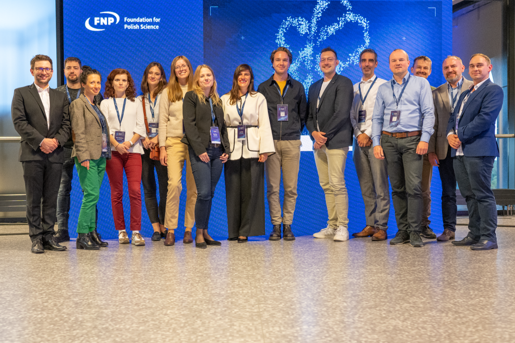
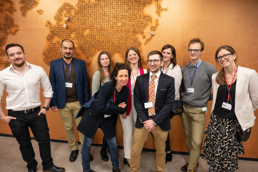
On February 29, 2024 the Foundation for Polish Science (FNP) announced that ICTER is among the winners of the first two calls for proposals in the “International Research Agendas” (IRAP) activity funded by the European Funds for Smart Economy Programme (FENG).

The IRAP FENG activity supports the establishment or development of specialized, world-leading research teams and organizations where scientific excellence and international research competitiveness can be achieved (source: FNP).

The focus of our research program, supported by this grant, is to contribute to advancements in the field of medical science. Specifically, we aim to develop new tools for safer and more effective surgical interventions, pioneer groundbreaking therapies for eye diseases, and create diagnostic methods that enhance prognosis and restore vision.

We extend our gratitude to the ICTER team, the Institute of Physical Chemistry, Polish Academy of Sciences, the International Scientific Committee, our partners, and collaborators, for their contributions to this achievement.

We look forward to leveraging this opportunity to further advance scientific excellence and international competitiveness in our eye research initiatives.

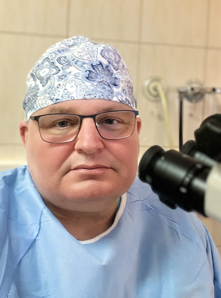
Photo: Karol Karnowski, PhD.

The „International Centre for Translational Eye Research” project is carried out within the MAB FENG action 02.01.of the Foundation for Polish Science co-financed by the European Union under the European Regional Development Fund, European Funds for Smart Economy, agreement no. FENG.02.01-IP.05-T005/23.

12.10.2023

Celebrating World Sight Day 2023 with the Foundation for Polish Science

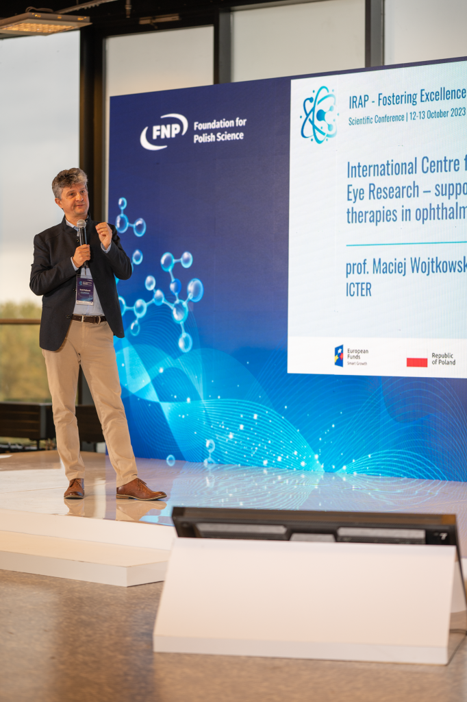
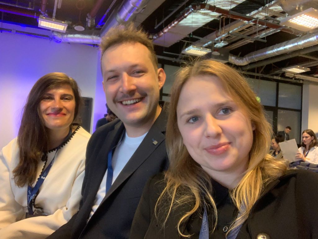
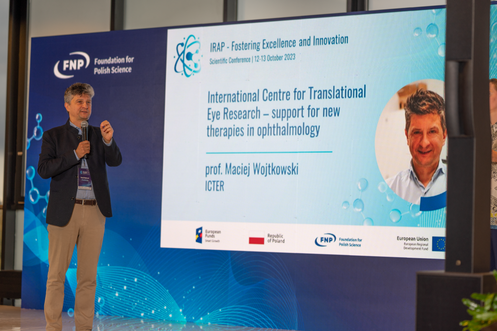
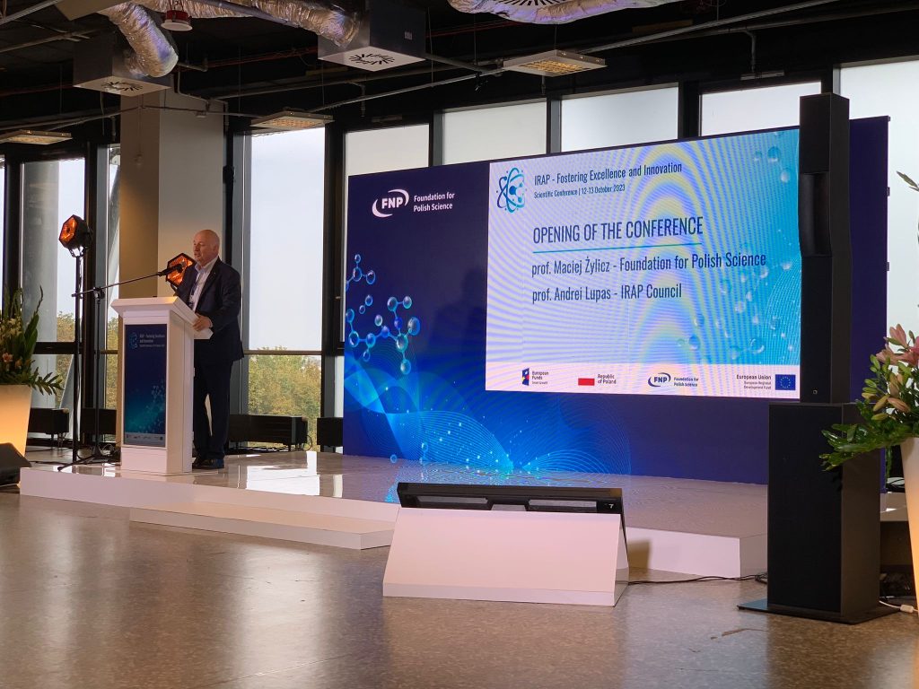
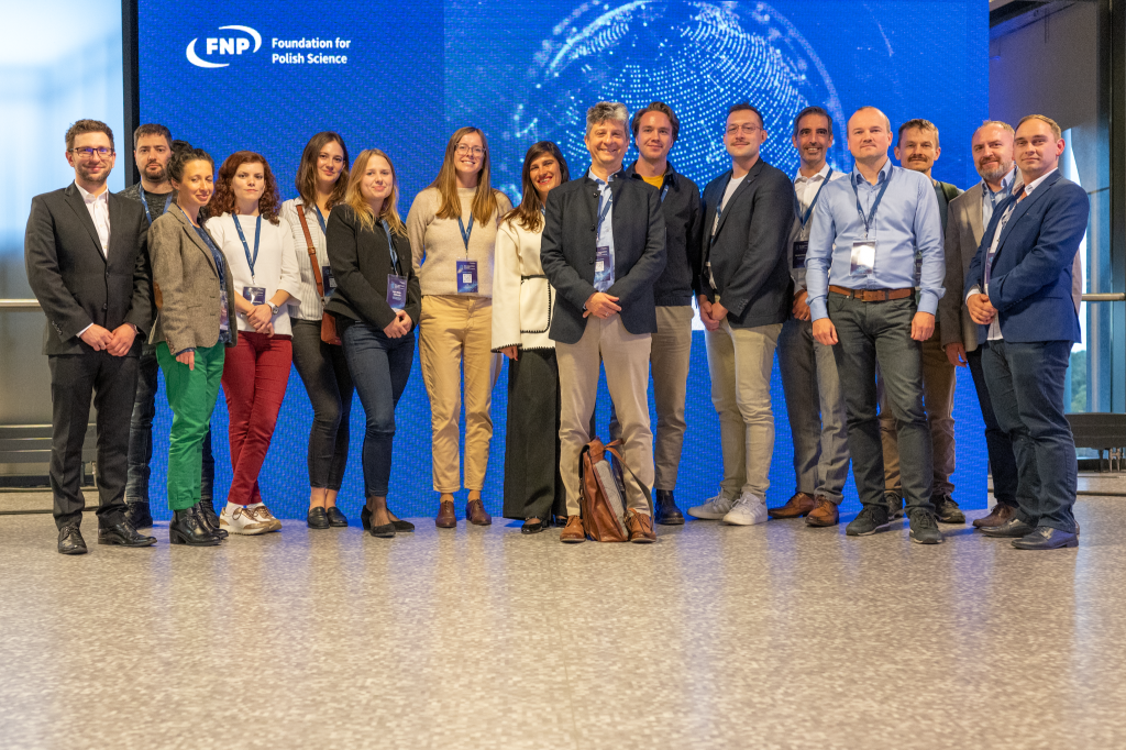
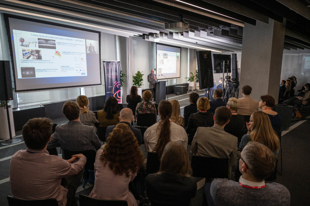
What better way to mark World Sight Day than to shine a light on the achievements of our dedicated team of researchers in advancing ophthalmic therapies? We can’t think of a more fitting occasion. On October 12, 2023, a strong delegation of our management and scientists embarked on a journey to represent ICTER at the “IRAP – Fostering Excellence and Innovation Conference” organized by the Foundation for Polish Science (FNP), the institution that played a pivotal role in establishing our centre. There, we presented our pioneering high-tech eye imaging methods, innovative biomedical solutions, and cutting-edge genetic therapies that have been instrumental in the realm of vision care and restoration.

Through an invited talk, Prof. Maciej Wojtkowski, who serves as ICTER’s Chair, summarized our International Research Agenda (IRAP) programme, highlighting our institution’s role in supporting new therapies in ophthalmology and promoting its achievements “as an example of excellence and innovation” (source: FNP). Prof. Maciej Wojtkowski is the IRAP laureate for the creation of ICTER (International Centre for Translational Eye Research), a centre that has received funding from the FNP under the Smart Growth Operational Programme.

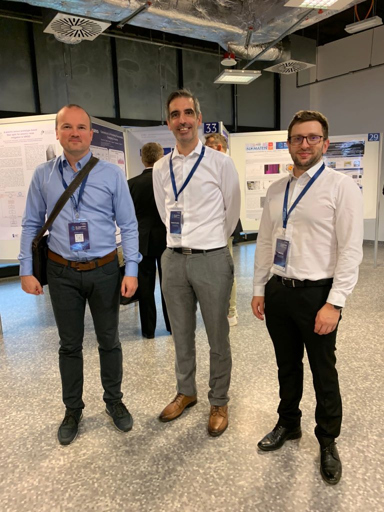
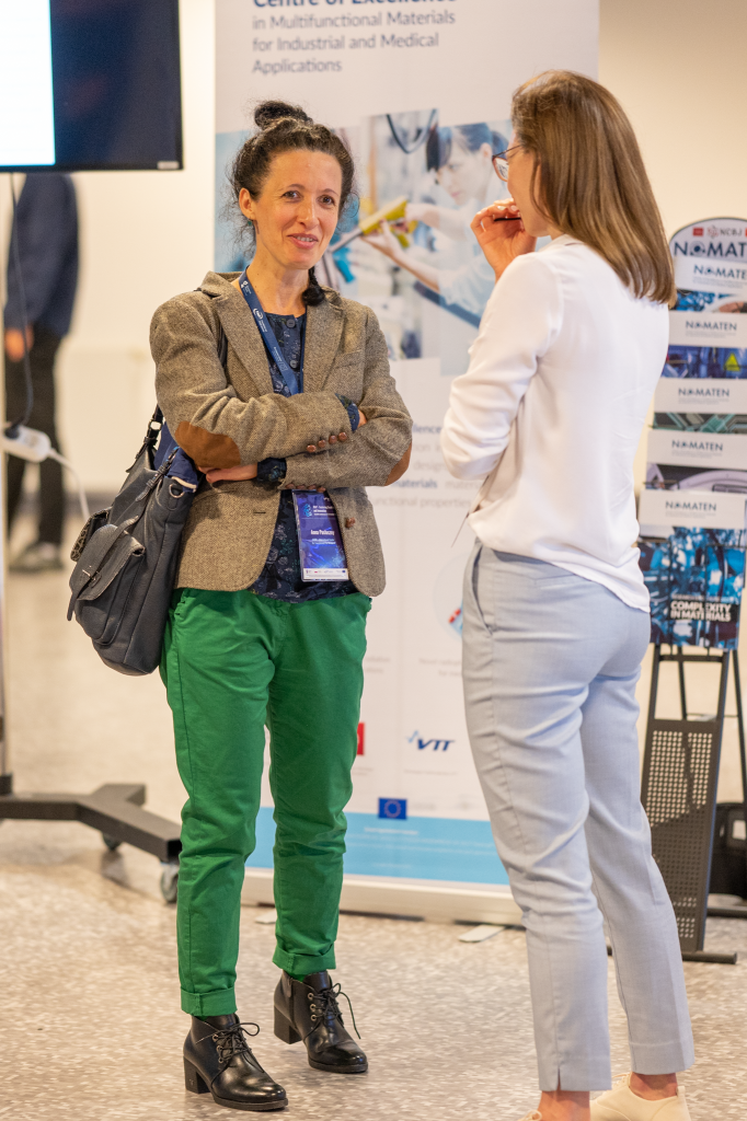
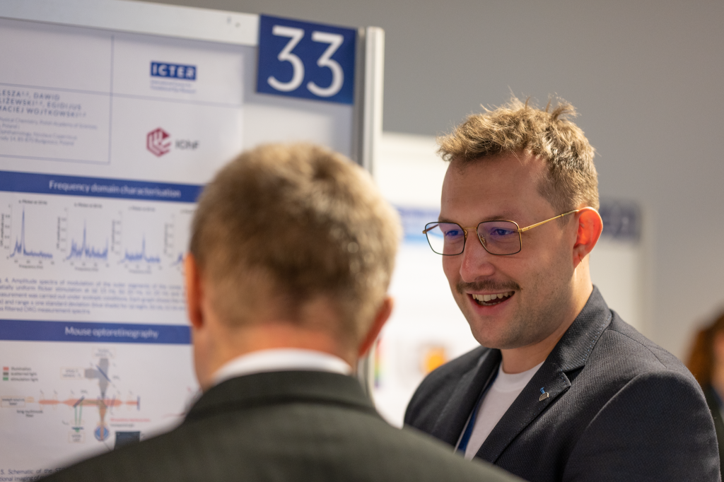
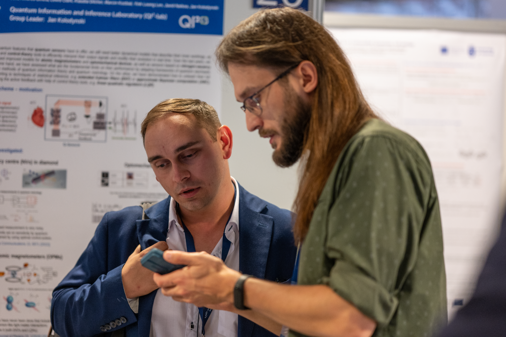
Our Principal Investigators were actively engaged at the conference, passionately showcasing the groundbreaking work undertaken in our labs through an array of informative posters and engaging direct discussions with other IRAP laureates, including directors, leaders, and research group members, who have received funding from the Foundation under the Smart Growth Operational Programme. They also interacted with the broader IRAP environment, including the IRAP Council, International Research Committee, and representatives from our business partners. Throughout the conference, we proudly presented cutting-edge research across a wide spectrum of fields, including Medical Physics, Biochemistry, Instrumentation Engineering, Pharmaceutical Sciences, Ophthalmology, Biomedical Engineering, and Ophthalmic Biology. Our expertise was exemplified through a series of scientific posters that showcased our strengths in Optical Instrumentation, Electrophysiological Data Analyses, Structural Biology, Bioinformatics, and the Design of Imaging Devices. These innovative areas represent our commitment to advancing eye care and revolutionizing the field of ophthalmology.

The ICTER Board was strongly represented by Anna Pawlus, our Managing Director, and the Deputy Director for Intellectual Property, Dr. Łukasz Kornaszewski. Our scientific community was also out in full force. Prof. Maciej Wojtkowski, was at the forefront, accompanied by Dr. Marta Mikuła-Zdańkowska and PhD student Piotr Wegrzyn, all from the Physical Optics and Biophotonics group. Dr. Marcin Tabaka, who leads the Computational Genomics group, was there with his team members Dr. Stefania Robakiewicz, and PhD student Piotr Rutkowski. Dr. Andrzej Foik, the leader of the Ophthalmic Biology group, was joined by his team members, including Dr. Anna Posłuszny, Dr. Katarzyna Kordecka, and Dr. Jagoda Płaczkiewicz. Dr. Humberto Fernandes, who leads the Integrated Structural Biology group, was accompanied by Luca Gesa, Nelam Kumar, Dr. Sathi Goswami, and Łukasz Olejnik. Furthermore, senior researcher Dr. Karol Karnowski, representing the Image-guided Devices for Ophthalmic Care group, was also part of our delegation.

Throughout the conference, we proudly presented cutting-edge research across a wide spectrum of fields, including Medical Physics, Biochemistry, Instrumentation Engineering, Pharmaceutical Sciences, Ophthalmology, Biomedical Engineering, and Ophthalmic Biology. Our expertise was exemplified through a series of scientific posters that showcased our strengths in Optical Instrumentation, Electrophysiological Data Analyses, Structural Biology, Bioinformatics, and the Design of Imaging Devices. These innovative areas represent our commitment to advancing eye care and revolutionizing the field of ophthalmology.

The full “IRAP – Fostering Excellence and Innovation Conference” transmission is available here.

Website of the event: IRAP – Fostering Excellence and Innovation Scientific conference – 12-13th October 2023 (irapconference.pl).

Photos: Dr. Karol Karnowski

Text: Dr. Anna Przybyło-Józefowicz

16.08.2023

A new paper by IDoc group researchers, international scientists and a spin-off company published in “Biomedical Optics Express”

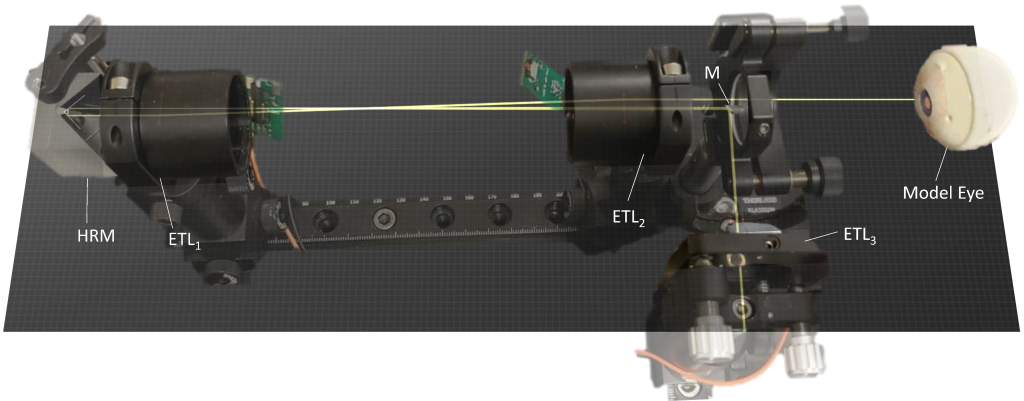
Whole-eye optical coherence tomography (OCT) imaging is a promising tool in ocular biometry for cataract surgery planning, glaucoma diagnostics and myopia progression studies. However, conventional OCT systems are set up to perform either anterior or posterior eye segment scans and cannot easily switch between the two scan configurations without adding or exchanging optical components to account for the refraction of the eye’s optics. In this work, we present the design, optimization and experimental validation of a reconfigurable and low-cost optical beam scanner based on three electro-tunable lenses, capable of non-mechanically controlling the beam position, angle and focus. The proposed beam scanner reduces the complexity and cost of other whole-eye scanners and is well suited for 2-D ocular biometry. Additionally, with the added versatility of seamless scan reconfiguration, its use can be easily expanded to other ophthalmic applications and beyond.

Text: Dr. Andrea Curatolo – Principal Investigator in the IDoc group at ICTER.

Publication:

María Pilar Urizar, Enrique Gambra, Alberto de Castro, Álvaro de la Peña, Onur Cetinkaya, Susana Marcos, and Andrea Curatolo, “Optical beam scanner with reconfigurable non-mechanical control of beam position, angle, and focus for low-cost whole-eye OCT imaging,” Biomed. Opt. Express 14, 4468-4484 (2023)

Link: https://opg.optica.org/boe/fulltext.cfm?uri=boe-14-9-4468&id=535917

23.06.2023

“Research conducted at the ICTER is not art for art’s sake. They improve ophthalmology and save patients’ lives” – interview with Dr. Piotr Chaniecki, Ophthalmic Surgeon

Ophthalmology is one of the fastest-developing fields of medicine. This is only possible by improving existing procedures and developing new eye treatment methods. We discuss the importance of the continuous development of ophthalmic techniques with Dr. Piotr Chaniecki.

What is the most crucial aspect of ophthalmology for you?

PC: Ophthalmology relies on technology. The most significant advancements in this field occurred after developing diagnostic devices and surgical techniques. The level and improvement of technology directly influence the precision of procedures and the effectiveness of direct diagnosis. The International Center for Translational Eye Research (ICTER) is focused on developing such devices. I see tremendous potential in creating new tools for doctors that will contribute to better and faster diagnoses.

As seen in Western clinics, ophthalmology in Poland is developing rapidly, but we still have a long way to go regarding technological advancement.

Why is the lack of specialized research being conducted in Poland that could help patients?

There is still much to be done. We are not lacking specialists, and I take pride in having trained several ophthalmologists, surgeons, and diagnosticians who now work as independent and excellent doctors in Polish clinics. In Poland, I observe a kind of stratification, with some places offering diagnostics and treatment at the highest global level while others require significant investment. Money is, of course, a problem, but not the only one – there is a lot of equipment in Polish facilities that is not always fully utilized. What is the reason for this? I can only speculate that it is due to a lack of ideas about how the equipment can be used for research, or perhaps it is due to a persistence in established procedures and routines. What I sometimes notice in conversations with doctors, including those working in academia, is a reluctance to change and challenge the status quo – if a diagnostic method works, why change it? If we can make a diagnosis based on average-quality results, why bother striving for more? Additionally, the entire system of training doctors requires many changes.

I can’t entirely agree with such an approach, which is one of the reasons I decided to collaborate with ICTER, as it holds great potential for the benefit of patients.

Dr. Piotr Chaniecki

From a clinical perspective, what equipment developed at ICTER is the most important?

My research shows many devices with enormous potential to improve surgical procedures. I firmly believe that some of them will be “milestones in global ophthalmology.” This is not art for art’s sake. Better equipment and technology mean better diagnostics and increased patient safety during surgical procedures. I’m referring to the possibility of reducing the number of complications in surgical techniques and increasing the accuracy of diagnoses. As an experienced ophthalmologist who performs procedures according to the highest standards, I know the criteria will be even more demanding.

What are the numerical occurrences of complications in your practice?

Complications are a particularly challenging topic for every doctor. Every active surgeon encounters complications, so it is true what they say, “those who don’t operate don’t have complications.” Complications can be considered statistically, but one must approach the numbers cautiously. Even Mark Twain wrote about statistics, stating there are three kinds of lies: lies, damn lies, and statistics.

When looking at complications numerically, one would need to consider a specific procedure, such as cataract surgery. Here, sources provide values ranging from 0.3% to 15% of cases, depending on the complexity of each case. I consider complications as lessons from which I continually learn. My statistics regarding complications are within the lower range of the statistical scale.

Congratulations.

This largely depends on accuracy, which is also influenced by technology. Technology developed at ICTER will undoubtedly contribute to reducing the number of complications during surgical procedures. Another area where I see tremendous potential is diagnostics. Advanced technology will certainly increase the accuracy of diagnoses and allow us to view a given pathology from a broader perspective. Wanting to cure a patient is not enough; we must first know what to treat.

How many cataract removal surgeries with intraocular lens implantation are performed in Poland?

In Poland, approximately 300,000 such surgeries are performed annually. Worldwide, around 20 million lens implantation procedures are carried out. These numbers have fluctuated significantly over the past three years due to COVID and geopolitical circumstances.

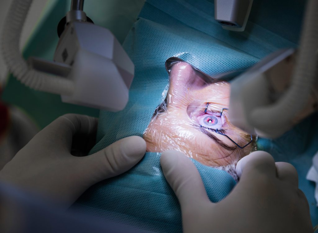
Ophthalmic surgery at an eye clinic

Gene therapy is another area being developed by ICTER. What prospects do you see there?

Gene therapy primarily offers a chance for visually impaired patients due to genetic disorders, such as those suffering from Leber congenital amaurosis (LCA). In individuals affected by LCA, the eye’s photoreceptors stop responding to light due to a mutation in the gene that codes for a protein essential in the visual process. Total blindness occurs around the age of 20. Research on gene therapy to remove or alleviate LCA symptoms has been ongoing for almost 15 years, and a viable treatment may soon be available. It is research institutes like ICTER that enable such progress.

Does gene therapy have a chance to become established in Polish medicine in the next few years?

We need to approach this topic realistically. Bringing a drug to market costs hundreds of millions of dollars. Research at each stage, including clinical trials, animal models, healthy volunteers, and patients, takes significant time. We are talking about a period of 5-10 years.

In addition to the research you are currently involved in with our scientists, focusing on patients with multiple sclerosis, do you plan to expand our collaboration to include patients with other conditions?

Indeed, in the next stage, we could involve age-related macular degeneration (AMD) patients. I see potential in diagnosing, monitoring disease progression, and assessing treatment effectiveness. Existing devices allow for structural imaging, which shows anatomical changes in different layers of the eye. Still, they do not provide functional imaging, meaning we cannot determine the state of crucial substances involved in vision biochemistry. Therefore, sometimes successful surgery does not result in improved vision for the patient. Such situations could be avoided if we knew beforehand whether the part we intend to repair is functioning. And this is where I see enormous potential in collaborating with the International Center for Translational Eye Research.

We want to benefit from your experience in ophthalmic practice, as it can help us refine the equipment we are developing. Do you have any guidance for us at this time?

First and foremost, for any device to be introduced into medical offices and operating rooms, it must be practical and user-friendly. It is not about the simplicity of the design or the principle of operation— not everyone needs to know how something works. Many people need to be able to operate the device. ICTER has developed many devices, such as systems for assessing retinal receptor function, which, with the suitable “packaging,” could quickly be implemented in clinics. The key is to create appropriate software so that the equipment can be operated by technicians or doctors after brief training without the need for an engineer. The second aspect is ergonomics and comfort for the patients. Let’s not forget that most patients are elderly individuals who may have mobility issues, not to mention spending 20 minutes in an immobile position during an examination. Additionally, some procedures can be particularly frustrating for them, primarily when they must focus on a bright spot they cannot see due to diseased changes in the retina. My goal is to present the clinical perspective to scientists.

Aside from my absolute satisfaction, our collaboration will benefit the patients the most. The fusion of technology, medicine, science, and practice always benefits all parties. The same will be confirmed in our case. I am eagerly looking forward to the results of this collaboration.

As are we.

Thank you for the conversation.

BIO

Piotr Chaniecki currently serves as the Chief Surgeon at the Prof. Zagórski Eye Surgery Center in Krakow. His professional background includes graduating from the Military Medical Academy in Łódź in 1996. He has also held the position of Head of the Clinical Ophthalmology Department at the 5th Military Clinical Hospital in Krakow and the Ophthalmology Department at the PCK Hospital in Gdynia.
His main areas of professional interest are anterior and posterior segment eye surgery, as well as conservative treatment of eye diseases. He is the author of a unique technique for intraocular lens exchange, which was recognized as the best surgical technique of 2019 by the American ophthalmic journal Cataract & Refractive Surgery Today. In 2016, he received the award for the best scientific paper titled “Composition of phacoemulsificated human lenses analyzed by infrared spectroscopy,” presented by the European Association for Vision and Eye Research.

———————————————————————————————————————————————————————————————-

The interview was conducted by a Postdoc researcher at ICTER, Dr. Michał Dąbrowski.

Proofreading: editor Marcin Powęska, MSc.

23.11.2022

Eyes well – dressed. We talk about the latest fashion trends in eyewear optics with the owner of the Studio Optyk optical store, Jarosław Bugaj

Nowadays, eyewear is a product that combines such diverse fields of knowledge and human activity as materials engineering, advanced digital technologies, ophthalmology and optical knowledge, precision craftsmanship, industrial design and art, and even luxury brand marketing. And in this view, they fit perfectly into the translational nature of an eye research centre like ICTER. Therefore, today we would like to introduce you to the topic of eyeglasses and eyewear fashion from the perspective of a person for whom their production, individual selection and repair are a personal passion and professional challenge.

Is it true that each of us will need at least a pair of eyeglasses during our lifetime? 

– Yes, it is true. It is inevitable. Sooner or later, even if we have not had to deal with glasses, at some point in our lives we might develop a young presbyopia, which is the loss of elasticity of the intraocular lens. Suddenly, we find that our hand needs to be longer to provide the proper distance to read the fine print. Then it is time to visit the ophthalmologist, measure the refraction, and check how much the natural lens is no longer efficient.

– So this means that we improve our correction by fitting lenses. But after all, we want to look well and fashionable, regardless of gender and age. 

– Yes, and then, in addition to the choice of lenses, we are faced with the choice of frames, which is not as easy as we thought. There are a lot of factors to consider and, above all, at the end of the day you have to like yourself and feel good in that eyewear. Often, we also want it to be in line with current trends. It used to be that glasses did not mean that much as today; they were rather considered a necessary evil. One had to wear them because he or she could not see properly without them. Nowadays, we also care about looking good in glasses and having something remarkable on our face; comfort and quality of craftmanship are also important. You could say that there are waves in eyewear optics; for example, there was a fashion for wire frames, then came the style for frames made of mass (plastic), and more recently transparent. Now it is all a bit mixed up. On top of that, each of us has individual preferences. Some people prefer strong frames that are clearly noticeable on the face and will mark their character, and others prefer softer, more subtle frames. It is essential that it is stylish and comfortable and that the patient feels good in the eyeglasses because it is a prosthesis of our sight, after all.

– Who sets the fashion trends in eyewear? 

– These days, major fashion houses mainly create fashion trends for ladies and gentlemen through their biggest, often exclusive, and most recognizable brands. In almost every seasonal collection, eyewear – mainly sunglasses but also corrective frames – is an integral part of the designs presented on the catwalk. Trends are also currently set by celebrities and influencers. Famous people, artists, and so-called stars often appear in eyeglasses, wearing increasingly exciting models, especially sunglasses, including those from the catwalks. Thus, they are arousing the interest of at least part of the public. We often have patients who ask for frames that a particular celebrity wears. This is interesting insofar as each face needs a slightly different frame. The one in which Christopher looks elegant and chic will not necessarily satisfy Charles, who also may feel uncomfortable wearing such frames. You also need to pay particular attention to individual preferences when choosing glasses.

 – Eyewear fashion is mainly shaped by the dictates of fashion houses. But other trends may impact on what we wear every day, too. A strong trend at the moment is ecology and sustainability. What does this trend look like in eyewear fashion? 

 – More and more manufacturers use recycled materials to make frames for sun and eyeglasses. We collaborate with a company that produces frames from raw materials from recycled ocean rubbish. More and more admixtures of wood and other natural materials appear in frames, which were initially poorly used due to their fragility and breakability. Nowadays, they are enriched with special accelerated plates, which make the luminaire more flexible and usable for longer. 

– Speaking of materials, what other materials besides those mentioned are used to create the frames?

Obviously, plastic. Today it is characterized by its enormous strength and lightness. These are usually thick frames, although lightweight, and also quite soft and therefore recommended especially for children. For this purpose, zylonite, or cellulose acetate – a hypoallergenic plastic – is used, and epoxy resin, which, when heated, is very malleable and easily adapts to the shape of the face. Metals such as surgical steel, aluminum, titanium, or beryllium are also used to manufacture frames. It all depends on the customer’s preference and wallet, as some frames, such as titanium, can be pretty expensive.

– Can you give a second life to your old eyeglasses? 

– Yes, some companies and foundations, also in Poland, collect used or unused frames and eyeglasses from opticians. In fact, I always have frames in my salon that I will not use, also for spare parts when repairing my customers’ glasses. From time to time, we pack them up and send them to the place where the frames are refurbished. Then a group of ophthalmologists and/or optometrists go to third countries and make glasses for people locally from these collected frames, so that they can be reused so that someone enjoys it. We walk around in a frame for a few years and it doesn’t wear out completely; after a few treatments, it can be refreshed. These are also helpful training materials for schools that train personnel of optical stores and optometrists, and this field of education has been growing rapidly in Poland for at least 10 years.

– Can you tell us about your adventure in optics?  

– My company is a family business. The business was developed by my father and he taught me the craft from childhood. I cannot imagine doing anything else. I am fascinated by glasses, optical technology, the selection of frames, how the eyeball is constructed, how the image forms on the retina. My dream is to go into optometry in the future. The current direction of optometry in Poland helps doctors to focus on the treatment of eye diseases, not only on the selection of vision correction. This is all the more so because technologies are becoming more and more advanced, the designs of the spectacle lenses themselves are basically changing from year to year, and the ophthalmologist is not necessarily aware of these design changes. The doctor focuses on the diseases, the final correction of the vision defect remains at the level of the personnel of optical store. When selecting glasses and frames, I prefer to check the prescription I get from the client, especially if it is for progressive or relaxed lenses, where there are progression channels or aberration zones. These are not big differences between the main prescription and my correction, often 0.5 dioptres or a slightly altered cylinder axis, but we are able to fine-tune this initial examination so as to squeeze the most out of the lens. Today’s lenses are more precise and require opticians to be more precise in their measurements as well. They are made using digital technology, where every 0.1 mm in fitting height or pupil distance or progression channel fit makes a huge difference to the patient. This significantly impacts patient’s comfort and adaptation to the new eyeglasses.


– Talking about technology, can you please tell us whether augmented reality is being used in the selection of frames, for example. Is this happening? 

– Yes, there are optical companies that are experimenting with it. The patient then stands in front of a device, a computer takes a scan of his or her face, then, based on an algorithm, calculates what frame would be optimal for that face and prints it in 3D. How this works in practice, I haven’t seen yet, but I’m curious to see if this selection – which is basically pure mathematics – will work in practice. After all, every face is different, and of course the computer can make very accurate scans, but whether this exact calculation will be good for a particular patient may always be a matter of dispute, as subjective issues come into play here, of one’s own judgement, which the machine is unable to assess. Of course, it is possible to write a program in which the patient can enter his or her preferences for such a setting, but there is no certainty that the final result will satisfy the patient. This already works in some places, but it is still not commercially applicable on a large scale. 

– Now, we often have several eyeglasses with different frames, especially when we have a visual impairment and do not want or cannot use contact lenses. We treat them a bit like a perfume or a watch. I am wearing this outfit today and I would like to wear matching eyeglasses.  

– Exactly, for me this is about sunglasses. I have a whole lot of them and I cannot get rid of any of them because I like them all. Today, glasses are an integral part of our image, whether we have a visual impairment or not. 

– While we are on the subject of functional glasses, what other types of glasses are still used in different areas of life? 

– There are, for example, sports glasses whose lenses are dedicated to golfers, pool players, runners, etc., who for various reasons cannot use contact lenses. Often these are special lenses, especially progressive lenses, designed to provide optimum functional comfort when playing specific sports. For example, for colliers who need to have full-spectrum vision. There are also safety glasses for people who work in difficult conditions – welders, turners – in which there is also the possibility of correction. An interesting example is ballistic goggles, designed mainly for advanced shooters over the age of 40, who encounter the problem inherent in all presbyopes, wanting to see the bow and target like in the good old days, but whose visual defect no longer allows them to do so. And this is also where dedicated glasses have their uses. 

– Where do you get from the eyewear that you offer? What are the leading countries in the production of frames and how does Poland rank against this?
 

– Poland ranks quite well, we have more and more domestic manufacturers of frames and the quality of these frames does not differ from their foreign competitors. The frames are really well made, high-quality materials are used in their production, and in terms of price they are definitely more friendly than foreign ones. I also have a wide range of frames from Italy and France in my showroom, as I love them for their design and often handmade, they are perfectly shaped and provide a high wearing quality. I also appreciate frames from Spain, which explode with colour and bold, modern styling, and are very lightweight and fun to wear. Polish frames are brilliantly made, but the design itself still needs work, and in this respect, we often take inspiration from foreign manufacturers. 

– How do you see the future of the eyewear industry?
 

– Since I have been in eyewear optics for more than 20 years, the industry has undergone a real revolution. Changes are happening right before our eyes, and they are being set by lifestyle, as customers’ needs are changing along with lifestyle changes. Hence the incredible popularity of sunglasses, which, in addition to their protective function, are actually a fashion accessory, new ideas from frame manufacturers for shapes, overlays, striking temples – everything that allows you to stand out from the crowd, emphasize your individuality and often also your material status. When it comes to design, many frame models are making a comeback. It could be said that every type of frame will have its time, albeit in a new guise. Also, material technology allows for incomparably more in design than before. 

Modern software on the ground of optical services is also entering the salons, including biometrics and VR technologies, which make it possible to acquire the extensive amount of data necessary to make individualized lenses optimally tailored to the needs of the individual patient. Such software is also being developed in Poland. In this respect, we are collaborating with Szajna in Gdynia, which is a manufacturer of progressive lenses and offers a VR diagnostic device to track the behaviour of the eye in real time with different accommodation and vision conditions. The data thus acquired provides additional information about the patient’s eye behaviour under different conditions and enables the optimal selection of progressive lenses. 

The future is happening today, and the optical industry itself has great potential for growth, not least because of the ever-increasing number of people requiring vision correction at different stages of life. I follow the latest eyewear trends with curiosity and attention in order to be able to provide my customers with a high-quality product that fully satisfies them medically, functionally and aesthetically.  

– Thank you very much for meeting me and I wish you the best of luck in the further development of your business!
 

The interview was written by Joanna Kartasiewicz, Research Funding Manager
, in collaboration with Jarosław Bugaj, owner of Studio Optyk Optical Store in Wolomin, near Warsaw. https://www.facebook.com/studiooptykwolomin/ 

Special thanks to Szajna company for the opportunity of testing their VR solution.
13.10.2022

Fascinated with the eye: Prof. Marco Ruggeri translates clinical needs into research, new ophthalmic technologies, and patents

On September 23, 2022, Prof. Marco Ruggeri of the Bascom Palmer Eye Institute visited our centre. His area of expertise includes instrumentation and quantitative imaging technologies for diagnostic and surgical applications in ophthalmology. Having a signed letter of intent with the Bascom Palmer Eye Institute, we discussed potential cooperation looking for joint projects to pursue, especially in the field of ophthalmic procedures. Our scientists Dr. Andrea Curatolo, Dr. Karol Karnowski, Dr. Slawomir Tomczewski, and Marcin Marzejon, M.D., gave Prof. Ruggeri a tour of the laboratories and discussed current research. Prof. Wojtkowski also met with the guest to talk about future projects. During the visit, Prof. Ruggeri gave an interview to our Communications and PR department about the popularization and dissemination of science in the United States and his approaches to promoting research and reaching the widest public with expert knowledge in the field of eye health and new ophthalmic technologies.

Interview with prof. Marco Ruggeri

Please tell us how your specialization translates into improvements in the state of expertise and excellence in vision research.

I work within several niches. First, we want to improve vision in old age so that people can preserve their vision quality later in their life. We first seek to understand why we lose our ability to focus on things up close with age by a condition known as presbyopia. To do so, we are studying the mechanics of accommodation, which is the autofocusing system of the human eye. This is the key part of the process as if we do not know how it works, we will not be able to fix it. We need to find out why we lose this ability as we age so we can counteract it. Since my specialty is optics and imaging, the way I do this is by visualizing and analyzing with our imaging technology what happens inside the eye in real life when we look at near objects and how that changes as we age. We also use this technology to assess the efficacy of the existing procedure to correct this condition, which is important as it provides feedback to manufacturers so that they can improve their products.

I also work on imaging technology for the early detection of eye diseases such as for example, keratoconus. This is important because, with our technology, clinicians will be able to act early and manage the condition in time to maximally preserve vision in patients. But there is more to it because these tools that we develop also provide clinicians with a way to understand whether the current therapies that they are using are effective or not, therefore improving the management of the disease.

As investigators working in translational research, our goal is to move basic science discoveries and technologies more quickly and efficiently into practice. Our vision research center is the ideal place to do so because we are literally located across the street from the hospital of Bascom Palmer Eye Institute, which is one of the largest in the nation. Our approach is to talk to clinicians and identify what the real clinical needs are, and then find a solution. We ask them what scientific discoveries would be game-changers in the field of ophthalmology and would make their life easier, and their feedback is worth focusing on. For example, our institute holds clinical grand rounds every Thursday morning where ophthalmologists confer on complex clinical cases that they discuss by listing different approaches to a given disease or injury. This is one of the best ways to understand what the clinical needs are. You just go there, listen, look at what they are doing, keep quiet, take notes, get ideas and talk to them. I have been doing this for years, and by now, I know most of the ophthalmologists at my hospital quite well. Some of these clinicians eventually became friends. I text them when I need their feedback on a research project, and they text me when they have a new clinical need. I realize this may not be a conventional way of setting scientific priorities, but for me, it proved to be extremely effective. And it has an additional benefit as it is an excellent way to disseminate my scientific work. I also send ophthalmologists my publications, presentations of my scientific work, and share with them the knowledge I explore, primarily driven by a grassroots clinical need.
To recap the life cycle of my work, I first look at a clinical need, and when I identify a meaningful project, I apply for funding to implement it. This is done by preparing a grant application together with a clinician. From the application submission to the funding of large multimillion-dollar from federal entities such as the National Institute of Health takes years, so it is important to be disciplined and act early. Once we receive the funding, I conduct joint research with the ophthalmologists, and the pathway is usually the same; we develop instrumentation and methods, we go into clinical studies on patients and see how it can affect clinical practice. The ultimate goal is to benefit the eye care of patients, so when we reach the end of a research project, and the technology is developed, we start approaching companies to see if they are willing to commercialize our technology bringing it to fruition for patients.

How did your adventure with optical imaging begin, and why did you choose this particular field?

It first started with the eye, even before optical imaging. The eye is a very fascinating part of the body from many points of view. It encompasses mechanical and optical functions, it converts light into electrical signals that travel to the brain and can be used as a window to the rest of the body. I got involved in eye research in Italy during my master’s degree thesis project in electrical engineering – an optical sensor to monitor the glucose concentration in the eye as a potential means of assessing blood glucose. Instead of detecting glucose concentration in the blood, the goal was to measure it non-invasively through the anterior chamber of the eye using an optical technique named polarimetry. That is how I got interested in eye research, but at that time, it was not imaging yet. After graduating, I looked for opportunities to work abroad in the field of measurement technologies applied to eye research. I then found a position as a research associate in the team at Bascom Palmer Eye Institute, developing one of the first implementations of high-resolution OCT retinal imaging for studying the human retina and the retina of small animal models of retinal diseases. It was during that time that I became familiar with the pioneering work of Prof. Wojtkowski on spectral domain OCT imaging. This year marks my seventeenth year at Bascom Palmer Eye Institute.

Are patients in the US aware that more accurate eye imaging methods lead to more effective therapies for eye diseases?

In my experience, not enough.

How do you disseminate your research results and publications?

I participated in the National Alliance for Eye and Vision Research, an organization that promotes advocacy and public education for the eye and vision research sponsored by the National Institute of Health and other federal agencies in the US. Every year they select a few researchers in the field of vision and train them to educate Congressional legislators, the media, and consumers about the value of eye and vision research. For example, we met with government policymakers and explained the importance of allocating taxpayers’ money to eye research and persuading them to promote more research funding for eyesight in the next bill. In the long run, this will save taxpayers money because the funded research will be spent to improve health care.

OCT imaging is a prime example of how technology can lead to significant savings of public funds, with an estimated of more than $10 billion in spending reductions over the last 15 years. The cost saving are the results of clinicians being able to provide more personalized eye care by using OCT to decide when prescription injection are needed to treat some forms of macular degeneration. Thanks to OCT, this process has been optimized by reducing the number of injections needed as well as complication and discomfort to patients.

As for the general population, there are not many channels to disseminate our research and stress its importance, but when it comes to popularizing science, I try to use the same simple language and message as for policy makers, showing the benefits of research applied in ophthalmology. Working in a hospital, I have the great opportunity to explain this directly to patients when they take part to our clinical studies. Other channels to reach the wider public are social media, such as Instagram, LinkedIn, Facebook.

What are the activities of the Bascom Palmer Eye Institute towards the promotion and PR of eye research and science?

Our communications and marketing department regularly publishes a magazine named “Images“ which focuses on the medical and scientific advances at our institution. For example, you can read how our doctors and scientists lead the fight against macular degeneration and how we help babies to see. We have also established a program with the local museum of science in Miami where scientists and clinicians from our institution organize evening seminars to educate the public to our research. Besides that, Bascom Palmer has official channels on social media too, and we are encouraged to work with them to promote our work directly on our institution profiles.

In your opinion, what is the best formula for bringing the nature, importance, and essence of the work of a scientist engaged in eye research to a wider public?

In general, scientists are more comfortable with the conventional and formal ways to disseminate research, such as publications in scientific journals, seminars, and presentations at conferences.  While this is important to passing on the benefits of our research to other researchers and professional practitioners, it has a limited outreach for the wider community. The newer generation of scientists is generally doing a better job at promoting the importance of their research on informal channels like social media platforms. Having a marketing department is a great tool for informing the public about research findings. As I explained before, direct contact with patients is helpful. Visits to schools are also a good way to introduce young people to science and get them used to the importance of scientific research. Popular science articles can also be published in the mainstream press, or events can be organized with the local museums for this purpose.

Do you notice any differences in the American and European approaches to science PR, and if so, what are they?

Europeans have made a great effort to promote their research; for example, we observe that scientists are encouraged to have their own laboratory websites or social media accounts. In the US, the promotion of scientists’ work is usually handled by the university’s communications departments. Europe also has great promotional mechanisms in place; for example, when receiving a grant, you are encouraged to advertise your research on a Twitter account. In the US, a special marketing department works for you; they are always looking for news, but we are not pressured, and it is only up to us how much we use their resources to make ourselves known to a wider audience.

What is your greatest professional goal in serving the public?

Generating solutions to improve eye care. The overriding sense of my work is to bring improvements to patients’ vision, ideally moving from research to commercial technology. My dream is that one day people in need can benefit from the technology I have developed.

What are your impressions of Poland and cooperation with Polish scientists so far?

I visited Poland in September of this year for the first time. My impression is that the Polish government is greatly investing significant amounts of resources and money in research. I see that scientific units have access to many grants and other sources of research funding. The cutting-edge technology developed by your centre and other institutions suggests that the level of education is very advanced in your country. Taking part in various conferences where I met Polish scientists, I can confirm that they never failed to present top-notch research. In addition, you are very open and value cooperation. I strongly believe in collaboration among scientists, and I consider that global research should evolve in the direction of international and interdisciplinary cooperation to unite forces and become complementary in what we do. This is the power of today’s science, enabled by modern technology and communication tools.
Thank you very much for the interview and for your visit to ICTER, Professor Marco Ruggeri. We look forward to working with you and can’t wait to start joint scientific projects.

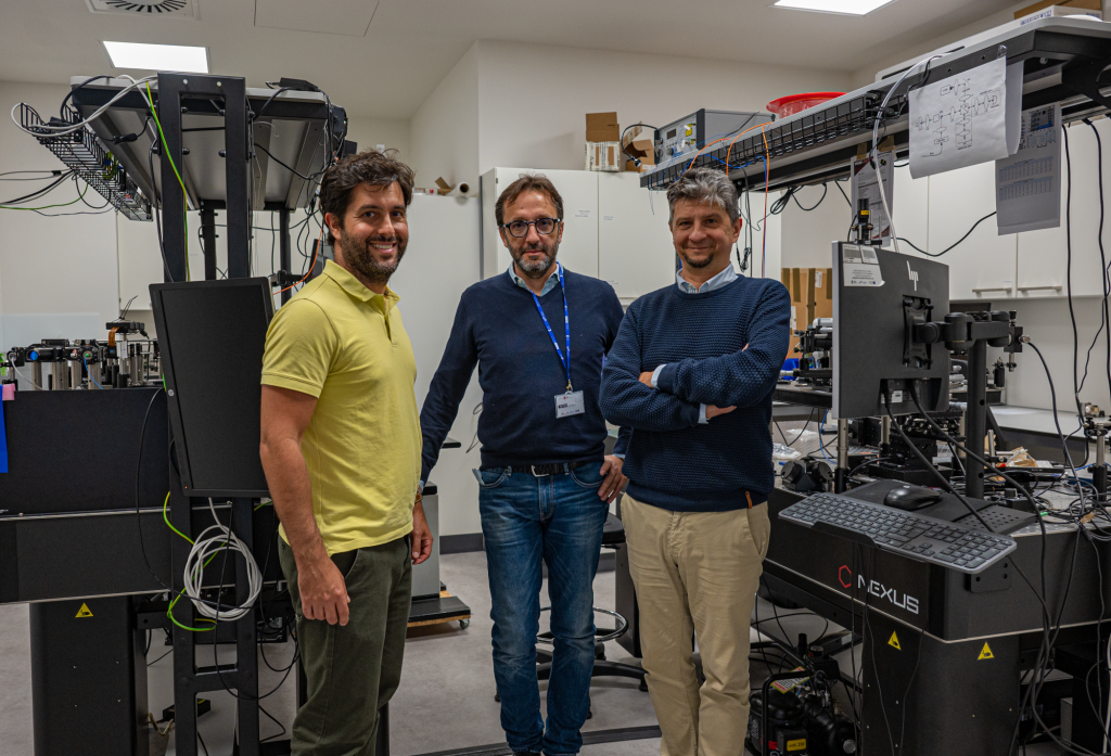
From the left to the right: Dr. Andrea Curatolo, Prof. Marco Ruggeri, and Prof. Maciej Wojtkowski.

Photo: Dr. Karol Karnowski.

The interview was conducted by the Communication and PR Manager, Dr. Anna Przybyło-Józefowicz.

10.03.2022

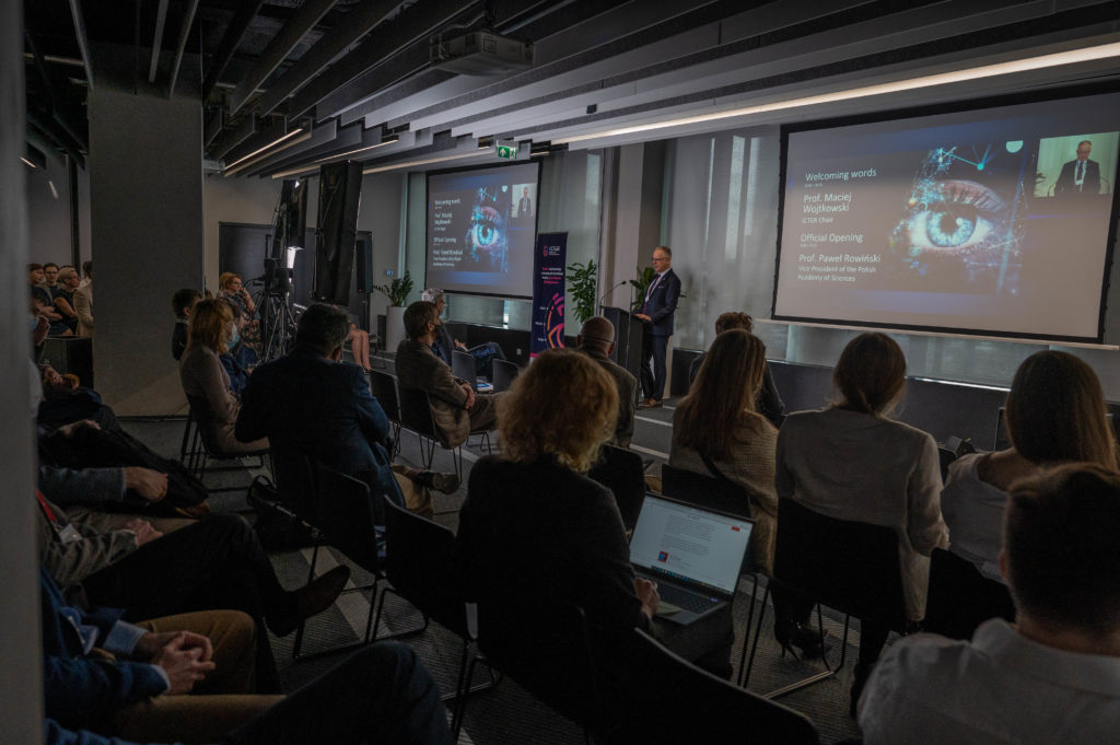
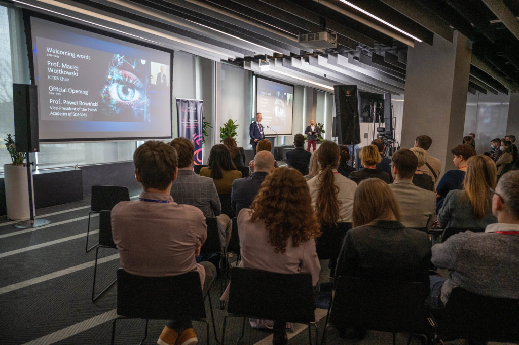
2022 ICTER ISC Meeting & Annual Review

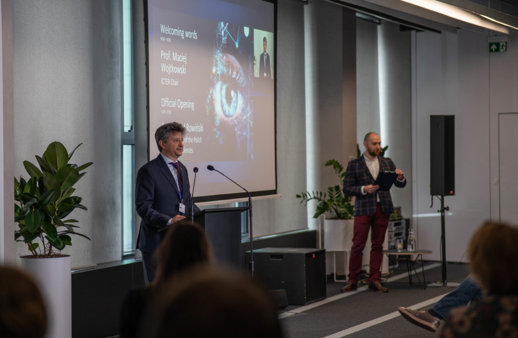
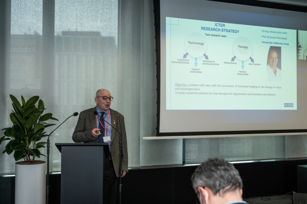
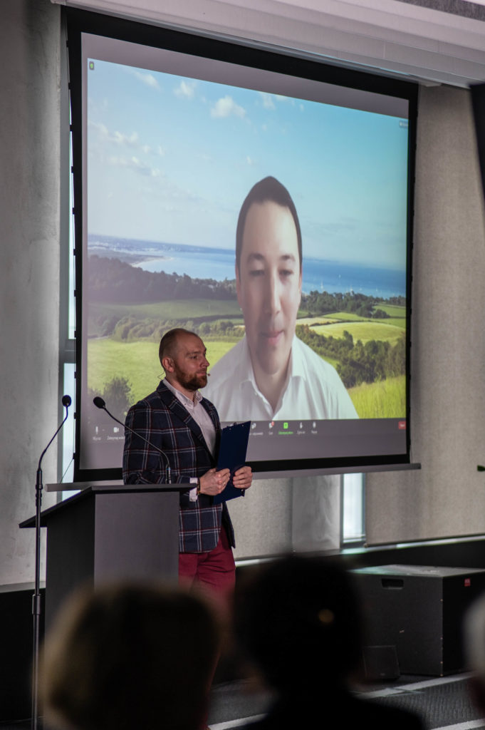
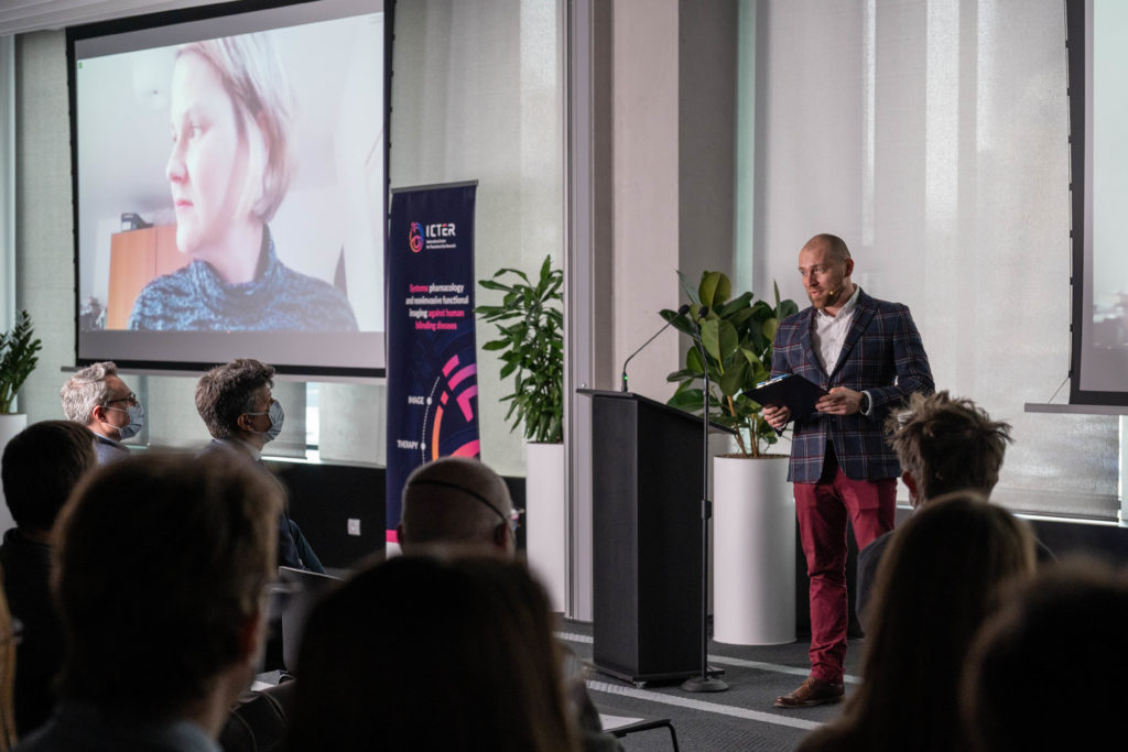
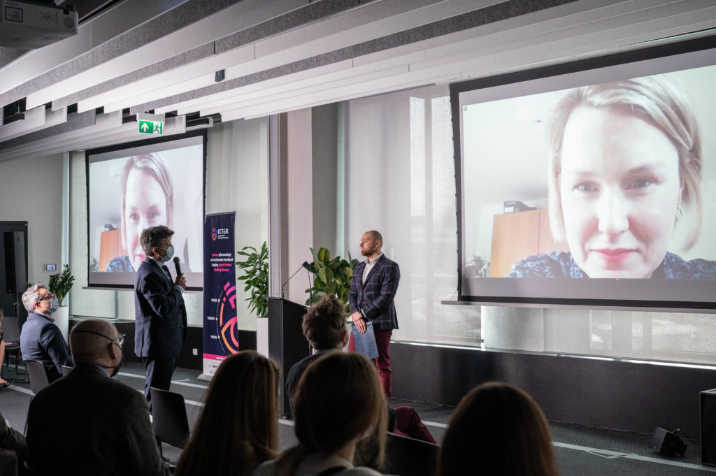
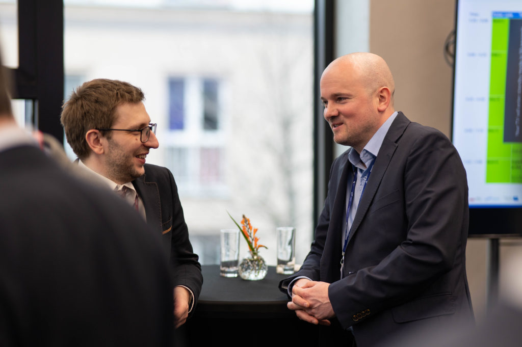
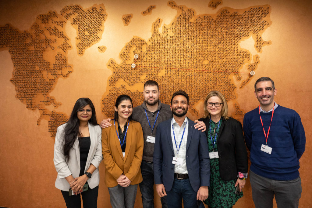
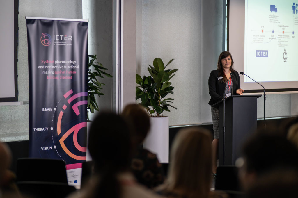
On March 9, 2022, we held the ICTER ISC 2022 Annual Meeting and Review. We met in hybrid mode, physically at Varso Place, downtown Warsaw, and online via Zoom. We summarized our scientific and research activities in 2021 for the experts of the International Scientific Committee, and presented strategic goals for our future research program. Invited guests from Poland and abroad delivered talks.

Dr Colin Chu from the Institute of Ophthalmology at University College London gave a lecture entitled: “Imaging Immune Responses in the Retina”. Dr Colin Chu is a Clinical Senior Research Fellow at the University of Bristol and Moorfields Eye Hospital, London. He has recently been awarded a Wellcome Trust Fellowship to start his lab at UCL Institute of Ophthalmology. He did his PhD at UCL before post-doctoral training with Prof Andrew Dick in Bristol and with Dr Ron Germain at the US National Institutes of Health. As a clinician-scientist and ophthalmologist his focus is on improving the care of patients with retinal diseases and studying fundamental immunology by applying in vivo and ex vivo imaging techniques to the eye.
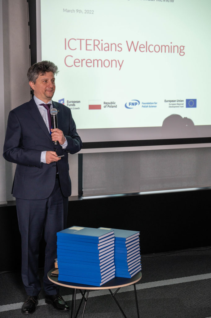
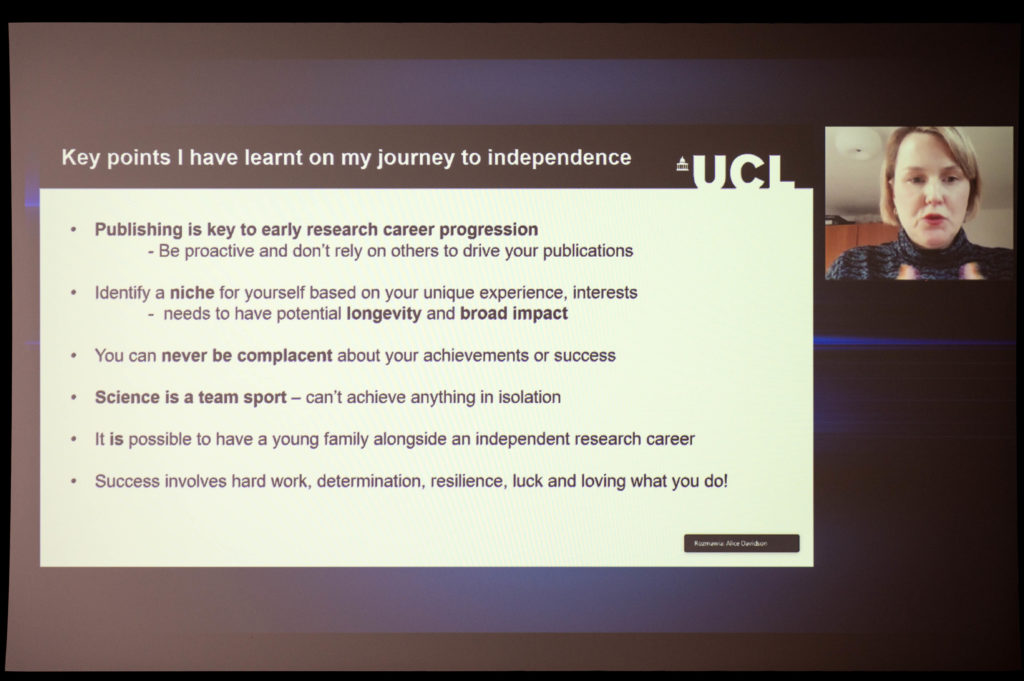
Dr Alice Davidson from the Institute of Ophthalmology at University College London gave a talk entitled “Corneal Endothelial Dystrophies: A Paradigm for Non-Coding and Repeat Expansion-Mediated Disease Mechanisms”. Associate Professor Alice Davidson is a UKRI Future Leader Fellow Professor at the University College London Institute of Ophthalmology (UCL IoO), London, UK. Dr Davidson started her scientific research career at the University of Manchester where she undertook a PhD in Molecular Genetics and Cell Biology (2006-2010), investigating the role of bestrophin-1 in ocular disease. She subsequently worked as a postdoctoral scientist at UCL IoO (2010-2015). In 2015 she was awarded a Fight for Sight Early Career Investigator Award to initiate her own independent research programme at UCL IoO and later a prestigious UKRI Future Leader Fellowship (2019) to further advance her inherited corneal disease research program.

Prof. Pearse Keane from Moorfields Eye Hospital in London gave a lecture entitled: “Artificial intelligence in ophthalmology – going from code to clinic”. Pearse Keane is Professor of Artificial Medical Intelligence at UCL Institute of Ophthalmology, and a consultant ophthalmologist at Moorfields Eye Hospital, London. He is originally from Ireland and received his medical degree from University College Dublin (UCD), graduating in 2002. In 2016, he initiated a formal collaboration between Moorfields Eye Hospital and Google DeepMind, with the aim of developing artificial intelligence (AI) algorithms for the earlier detection and treatment of retinal disease. In August 2018, the first results of this collaboration were published in the journal, Nature Medicine. In May 2020, he jointly led work, again published in Nature Medicine, to develop an early warning system for age-related macular degeneration (AMD), by far the commonest cause of blindness in many countries.
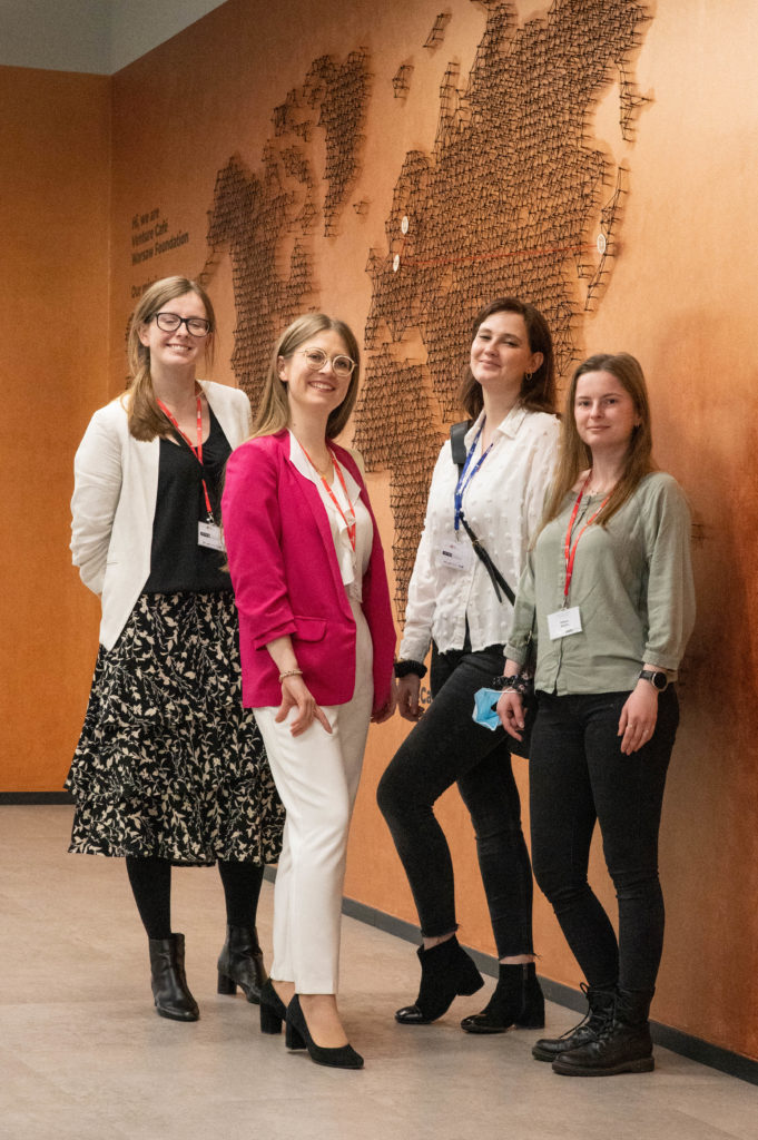

Prof. Krzysztof Palczewski of the University of California, Irvine gave a visionary lecture entitled: “Precise genome editing in the eye”. Prof. Palczewski is a co-founder of ICTER. He is a biochemist and a globally renown expert in biochemistry of vision – employed in the Medical School at the University of California Irvine, USA. Author of more than 500 scientific papers published in the leading magazines, including “Science,” “Nature” and “Molecular Cell” – filled over 10 patents and patent applications. His significant achievements include crystallizing and describing the structure and function of rhodopsin, as well as discovering the mechanisms leading to retinal degeneration and, in consequence, to vision loss.

At the meeting, representatives from the following authorities also attended and delivered presentations:

Polish Academy of Sciences: Vice President Prof. Paweł Rowiński,

Institute of Physical Chemistry of the Polish Academy of Sciences: Deputy Director for Scientific Affairs Dr. Adam Kubas, Professor of the Institute,

Foundation for Polish Science: President of the Board Prof. Maciej Żylicz.

All pictures were made by our senior researcher & photographer dr Karol Karnowski.

The master of ceremonies of this event was Marek Ziemba.