Worldwide, as many as 285 million people suffer from serious eye diseases or blindness. Unfortunately, most of them do not have access to modern methods of treatment, so help often comes too late. This situation could change with the advent of a very significant improvement to a diagnostic tool that has been utilized for three decades for detecting ocular pathology – Optical Coherence Tomography (OCT).
OCT is one of the most basic and accurate tests used in the diagnosis of eye diseases. It allows for a detailed view of individual eye structures, and thus enables the detection of macular diseases, diabetic changes of the retina, glaucoma, ocular tumors, and many other disorders. Unfortunately, the OCT method is flawed, because naturally occurring noise during the eye examination reduces the accuracy of imaging. A team of researchers at the International Center for Translational Eye Research (ICTER) set out to correct this problem, revolutionizing the OCT method by introducing spatio-temporal OCT Tomography (STOC-T).
Badania zostały przeprowadzone przez dr Edgidijusa Auksoriusa, dr Dawida Boryckiego, Piotra Węgrzyna i The research was conducted by Dr. Edgidijus Auksorius, Dr. David Borycki, Piotr Węgrzyn, and Prof. Maciej Wojtkowski of ICTER, and the results were published in the journal “Optics Letters” in a report entitled, “Multimode fiber as a tool to reduce cross talk in Fourier-domain full-field optical coherence tomography“.
How does the OCT examination work?
Owing to its high resolution the OCT method is one of the most frequently used ophthalmologic examinations. It is completely painless and safe – there are no medical contraindications to its use (the examination can be performed even with pregnant women). OCT works best in diagnosis of eye diseases such as progressive glaucoma, diabetic retinopathy, or age-related macular degeneration (AMD), which are the most common causes of central vision loss in older people. For example, in the early stages of AMD, single deposits or clusters of pigment and subtle atrophic changes are visible on the fundus via OCT; with the development of diabetes, changes in the microvascular structure of the retina are observed on OCT images. The OCT examination itself takes a few minutes. The patient sits in front of a special apparatus and must focus on a point indicated by the doctor, limiting blinking. The measuring head is set 2-3 cm from the eyeball, so there is no possibility that it has any contact with the patient’s eye. In most cases OCT examination does not require any special preparation – the patient can come alone by car. However, the interpretation of the results is complicated, so it should be carried out by an experienced ophthalmologist.
Biophysics of OCT
Zrozumienie podstaw fizycznych badania OCT nie jest łatwe. Technika ta pozwala na przeprowadzenie “Understanding the physical basis of OCT examination is not easy. This technique is equivalent to performing a noninvasive “optical biopsy” in real time to visualize the microstructure of the tissue and diagnose possible pathological changes. In optical tomography, all data about the structure of the object are obtained on the basis of the intensity of the interference signal (formed by the superposition of two laser beams). OCT optical tomography, now used in ophthalmology offices around the world, takes advantage of an interesting property of light called coherence, in the dimensions of time and/or space. Classical OCT uses partially coherent light sources (temporally, but not spatially coherent) – the detector measures the difference in optical paths between the mirror in the interferometer and successive layers of the sample object (eye).
Inside the interferometer is a special plate that divides the rays into two parts and records the interference of the ray reflected from the tissue structures and the incident ray. Knowing the differences of the optical paths, the position of the analyzed eye structures can be determined. The data are processed by a computer and then presented in the form of two-dimensional cross-sectional images (tomograms). Tissues are multi-component structures, which scatter light in different ways. Depending on the degree of reflection or absorption, a grayscale or color image is presented. Objects with the highest reflectance are seen in red or white, and those with the weakest signal appear as dark colors or dark gray. Tissues with intermediate reflectance values are present in yellow-green or shades of gray. OCT uses low-coherence interferometry, in which interference occurs at the micrometer level (through the use of superluminescent diodes or short-pulse lasers). Infrared radiation sources are usually used. Non-coherent light sources (e.g., halogen, LED, or incandescent lamps) cannot be used in classical OCT examination. A team of scientists from ICTER was the first in the world to combine the properties of light coherence in both time and space, which enables more accurate diagnostic images of the eye.
How can OCT be improved?
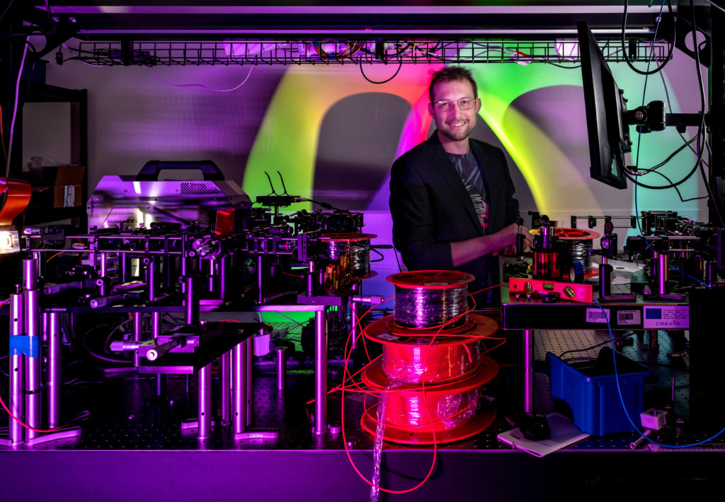
Spatio-Temporal Optical Coherence Tomography (STOC-T) is an especially effective tool for imaging the eye due to its speed and ability to acquire stable phase information over the entire visual field (unlike focused beam scanning). Until now, the main problem in using the OCT method has been noise (called speckle), which has made it difficult to accurately visualize the choroid, a vital part of the eye that supplies oxygen and nutrients to photoreceptors, and consequently is involved in the pathogenesis of many diseases. ICTER researchers found that using a multimode optical fiber of the appropriate length improves the imaging of the eye.
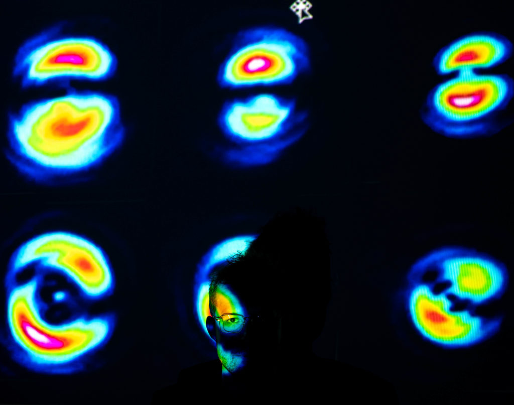
Multimode optical fiber emits several hundred unique spatial patterns (so-called transverse electromagnetic modes (TEM)) at its end in the cross-section of the beam. Until now, such devices have been used repeatedly to transmit data, but no one considered the fact that each of the spatial patterns come out of the several hundred meters of such optical fiber at different times. This time-dependence results in several hundred OCT images being captured during a single measurement; when added together the composite pattern reduces unwanted effects such as speckle noise in totally passive way. Applying this idea to OCT, a team of researchers at ICTER has developed a new way to control the optical phase of STOC-T to obtain high-resolution images of the retina and cornea in vivo. This method now allows much better cross-sectional images to be obtained from the choroidal layer under the retina, which previously was not possible.
OCT is one of the routine ophthalmic examinations used worldwide. Thanks to the improvements by the ICTER team, the advanced STOC-T technique will enable identification of changes in the eye at the cellular level, which will translate into better diagnoses and understanding of the onset and progression of various blinding diseases.
Author: Marcin Powęska
Photos taken by: Karol Karnowski


Publication
Title:
Authors:
Egidijus Auksorius, Dawid Borycki, Piotr Wegrzyn, Ieva Žičkienė, Karolis Adomavičius, Bartosz L. Sikorski, and Maciej Wojtkowski.
Journal:
Optics Letters Vol. 47,Issue 4, pp. 838-841(2022) DOI: https://doi.org/10.1364/OL.449498


