One of the primary initiatives within the IDoc Group focuses on the development of safer and more effective tools for eye surgery. This endeavor posed a distinct challenge for us, as it fell outside our traditional areas of expertise. Nevertheless, it is indeed a remarkable achievement that we have managed to assemble a team that, in under three years, has successfully amalgamated diverse expertise and advanced the project to its current stage. Our journey was marked by a gradual accumulation of knowledge and experience, ultimately culminating in the integration of all the components.
We are now pleased to inform you of the initial experiments where a robotic manipulator has been deployed to enhance manual ophthalmic surgical procedures. These innovations are complemented by the integration of Optical Coherence Tomography (OCT) images, meticulously aligned with the surgical tools’ tip precise position.
Another project the IDoc laboratory has been involved in goes to the very core of what the ICTER research centre aims to develop, that is methods and instrumentation to detect proper eye structure and function and their alteration in case of disease in an objective way. We did so in collaboration with the POB lab, by pioneering a technique called optoretinography. We are combining this with structural biology tools from the ISB lab for analysing the cellular machinery and its complex changes during the visual cycle in order to validate our hypotheses about what process the functional signal we measure originates from. To do so we are validating our functional imaging results with electrophysiology methods together with OBi laboratory.
It has been a challenging and ambitious project so far, but its collaborative nature made it all the more rewarding when not long ago we observed, in a repeatable way and for the first time, reduced functional responses from mice subject to temporal inhibition of vision, compared to their response only a couple of hours before the pharmacological treatment. We were able to objectively show with optoretinography that when a central protein (from the PDE family) involved in the phototransduction is inhibited, the mouse retinal photoreceptors, when exposed to a short flash of light, do not elongate nearly as much as they do when the mouse eye is fully functional. Measuring such a small physical change on photoreceptor length in vivo, as we are talking of only few tens of nanometers, can have a huge impact on vision science and ophthalmology by providing an objective functional test of visual ability and photoreceptor health. This in turns can speed up therapy selection and efficacy studies.
We look forward to upcoming results in this field.
Microendoscopes are the cornerstone of modern medical diagnostics – they allow us to see what we could not even describe two decades ago. The technology is constantly improving, with ICTER scientists contributing to the development of the probes.
Microendoscopes using fiber optics are becoming increasingly important imaging tools, but they have physical limitations. They are essential for applications that require a long working distance, high resolution, and a minimum probe diameter. The research paper titled “Superior imaging performance of all-fiber, two- focusing-element microendoscopes,” by Dr. Karol Karnowski of ICTER, Dr. Gavrielle Untracht of the Technical University of Denmark (DTU), Dr. Michael Hackmann of the University of Western Australia (UWA), Onur Cetinkaya of ICTER and Prof. David Sampson of the University of Surrey, sheds new light on modern microendoscopes. It is noteworthy that the research work started while the authors worked in the same research group at UWA.
In it, the researchers showed that endoscopic imaging probes, particularly those for so-called side viewing, combining fiber-optic (GRIN) and spherical lenses, offer excellent performance over the entire range of numerical apertures and open the way to a broader range of imaging applications. In the publication, the performance of endoscopic imaging probes is comparable to commonly used single focusing element probes.
Photo: Karol Karnowski
What are microendoscopes?
Miniature fiber-optic probes, or micro-endoscopes, allow imaging of tissue microstructures deep into the specimen or patient. Endoscopic Optical Coherence Tomography (OCT) is particularly promising. It is suitable for volumetric imaging of external tissues and the interior of organs (e.g., the upper respiratory tract, gastrointestinal tract, or lung tubules).
Three main ranges of fiber optic probes can be distinguished. Studies of large, hollow organs (such as those above the upper respiratory tract) require the largest imaging depth ranges (up to 15 mm or more from the probe surface), which can usually be achieved with low-resolution Gaussian beams (spot size in focus in the range of 30-100 μm). The intermediate resolution range (10-30 μm) is helpful for broader applications, such as imaging the esophagus, smaller airways, blood vessels, bladder, ovaries, or ear canal. The biggest challenge is obtaining beams with a resolution better than 10 μm, potentially helpful for animal model studies.
Photo: Karol Karnowski
When developing a probe, one must be mindful of design parameters’ trade-offs and their impact on imaging performance. Optical systems with a large numerical aperture (high resolution) tend to have a shorter working distance (WD). In addition, better resolution and longer working distance are more difficult to achieve as the probe diameter is reduced. This can be particularly problematic for side viewing probes – a greater minimum working distance is required compared to their forward imaging counterparts. Suppose the probe is encased in a catheter or needle. In that case, this increases the required minimum working distance – in many cases, this is the limiting factor in minimum achievable resolution or probe diameter.
It’s worth noting that engineers are usually interested in minimizing the probe diameter for reduced perturbation to the sample and patient comfort. A smaller probe means a more flexible catheter and, therefore, better tolerance of the test by the patient. Thus, one of the best solutions is using monolithic fiber optic probes, whose diameter is limited by the thickness of the optical fibers. Such probes are characterized by ease of fabrication, thanks to fiber-optic welding technology, which avoids the need for tedious alignment and bonding (usually gluing) of individual micro-optical components.
Photo: Karol Karnowski
Different types of microendoscopes
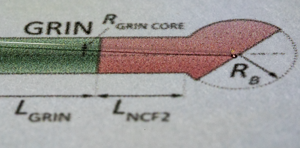
The most popular designs of fiber-optic imaging probes are those based on two types of focusing elements: GRIN fiber probes (GFP – GRIN fiber probes) and ball lens probes (BLP – ball lens probes). GRIN probes are easy to make, and their GRIN refractive power is not lost when the refractive index of the surrounding medium is close to that of the fiber used. Commercially available GRIN fibers limit achievable designs. High resolution is tough to achieve with GRIN fibers with small core diameters.
For lateral viewing probes, the curved surface of the fiber (and potentially the catheter) introduces distortion that can adversely affect imaging quality. Spherical BLP probes will not have this problem, but a sphere bigger than the fiber diameter is often required to achieve a resolution comparable to GFP probes. The focusing power of a BLP probe depends on the refractive index of the surrounding medium, which is an important issue when working in a medium with close or near biological samples.
One solution to improve the performance of probes is to use multiple light focusing elements, similar to the design of lenses with a long working distance. Studies have shown that combining numerous light-focusing elements provides better results for many imaging purposes. Probes with multiple focusing elements can achieve better resolution with smaller diameters while offering longer working distances without sacrificing resolution.
How do we improve the probes?
In their latest work, researchers led by Dr. Karnowski have shown that probes with two focusing elements using both GRIN segments and spherical lenses – called GRIN-ball-lens probes (GBLP) – significantly improve the performance of monolithic fiber optic probes. Their first modeling results have already been shown at conferences in 2018 and 2019. GBP probes were compared to the most commonly used GFP and BLP probes and showed performance benefits, especially for applications requiring longer operating distances, better resolution, and small size.
For intuitive visualization of probe performance, the researchers introduced a novel way to comprehensively present simulation results, especially useful when more than two variables are used. Analysis of the effect of GRIN fiber length and spherical lens size led to two interesting conclusions: for optimal results, the range of GRIN fiber length can be kept in the field of 0.25-0.4 pitch length (so-called pitch length); even if the working distance (WD) gain is not so significant for GBLP probes with high numerical aperture, the authors showed that the same or better performance in terms of working distance is achieved for a search with twice the diameter. Moreover, the novel GBLP probes offer higher resolution compared to BLP probes.
Photo: Bartłomiej A. Bałamut
The paper’s conclusion reads:
We have demonstrated the potential of GBLP probe design for applications with increased working distance, significant for lateral imaging probes, with a highly reduced impact of the refractive index of the probe’s environment and a significantly smaller size compared to BLP or GFP probes. These advantages make GBLP probes a tool worth considering for many imaging applications in biological and biomedical research, particularly for projects requiring micro endoscopes.
Note: The first results from “GRIN-ball-lens probes (GBLP)” modeling have already been shown at the 2018 and 2019 conferences:
– Karol Karnowski, Gavrielle R. Untracht, Michael J. Hackmann, Mingze Yang, Onur Cetinkaya, David D. Sampson, “Versatile, all-fiber, side viewing imaging probe for applications in catheter-based optical coherence tomography,” Photonics West, San Francisco, USA, Feb 2019, oral presentation;
– K. Karnowski, G. Untracht, M. Hackmann, M. Yang, O. Cetinkaya, and D. D. Sampson, “Versatile, monolithic imaging probes for catheter-based OCT,” 15th Conference on Optics Within Life Sciences, Rottnest Island, Australia, Nov. 2018, poster presentation.
The team responsible for these results started at the University of Western Australia (UWA), and work has now been completed within the following institutions: the Institute of Physical Chemistry, Polish Academy of Sciences and the University of Surrey, one of the authors only remaining at UWA.
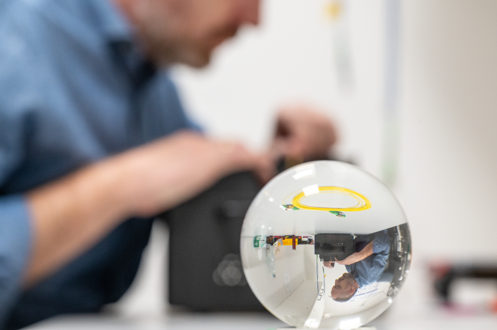
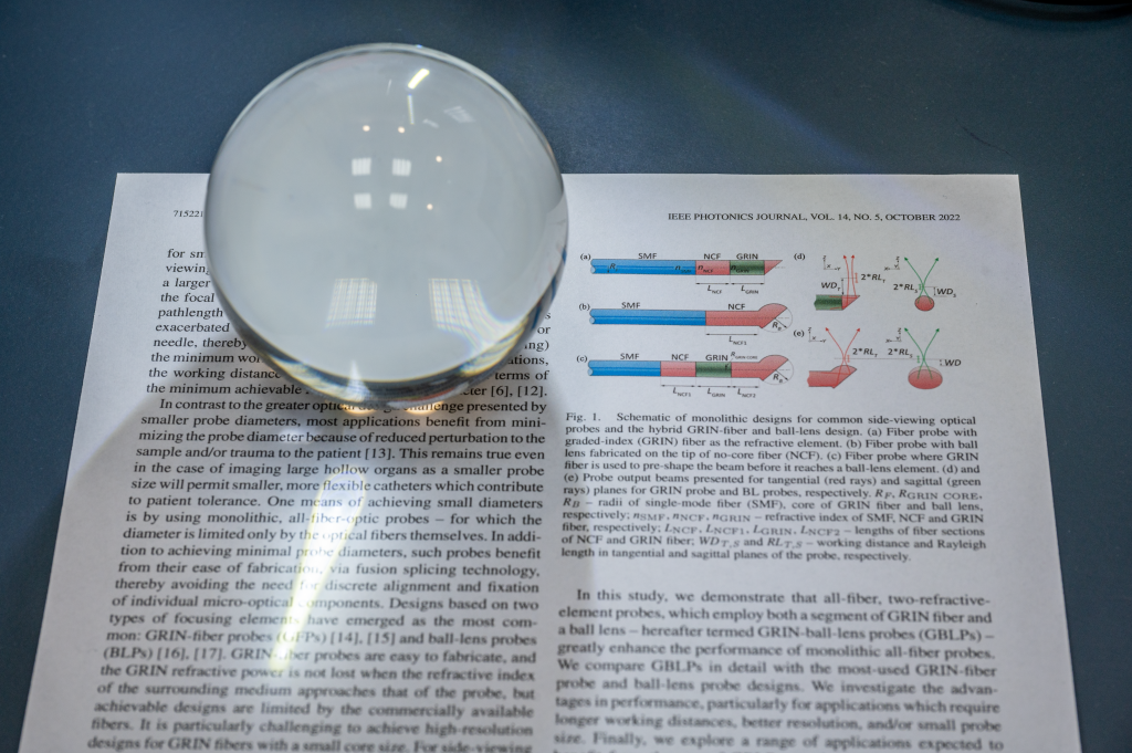
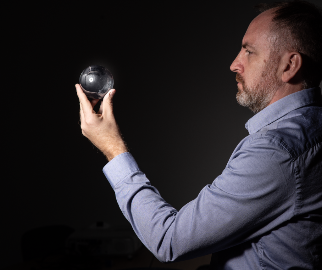
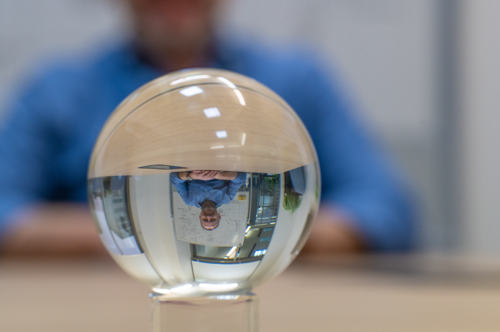
Photographers’ comment: One of the key components of the developed probes is a spherical surface on the tip of the fiber. In the photos, we used the imaging capabilities of such spherical elements (glass sphere).
Cited paper: K. Karnowski, G. Untracht, M. Hackmann, O. Cetinkaya and D. Sampson, “Superior Imaging Performance of All-Fiber, Two-Focusing-Element Microendoscopes,” in IEEE Photonics Journal, vol. 14, no. 5, pp. 1-10, Oct. 2022, Art no. 7152210, doi: 10.1109/JPHOT.2022.3203219.
Funding sources:
Polish National Agency for Academic Exchange (NAWA) through the Polish Returns Program
The main aim of our Institute is to revolutionize the diagnostics and treatment of eye diseases. Today, during World Sight Day 2021, special public awareness is raised on blindness and vision impairment as major international public health issues.
Want to explore more – visit World Sight Day website – https://www.iapb.org/world-sight-day/
#LoveYourEyes
What can we, individuals do on World Sight Day (and better on daily basis)?
encourage 50+family members and friends to have regular sight checks so potential diseases are diagnosed early,
if you spend hours in front of screens (laptops, phones, …) remember to make a small break every 20-30 minutes to look at a far distance (about 6 or more meters) for 20 seconds. This will provide some relax to our eyes,
you can also have a closer look at your diet. Antioxidant, vitamin and microelements reach products as yellow, orange and green veggies contain lutein that has beneficial effects for retina function. Choose omega-3 acid products (fat sea fish, legume and Italian nuts). Blueberries, pumpkins, apricot are good sources of antioxidants.
A decade ago, two scientists from our Institute – prof. Wojtkowski and dr Karnowski – published the world’s first air-puff Optical Coherence Tomography research [1]. The proposed method for direct measurements of apex corneal deformation was explored in several follow-up studies [2-4].
Over the last 4 years, prof. Wojtkowski and dr Karnowski lead locally (at the Institute of Physical Chemistry Polish Academy of Sciences) a group of researchers within the IMCUSTOMEYE – a 4-year project funded by the European Commission’s Horizon 2020 Programme under the Photonics 2017 KET topic. The IMCUSTOMEYE project focuses on the progress of the space of the air-puff OCT method towards three-dimensional measurements [5]. The ultimate goal is to enable the characterization of the ocular mechanical behavior in vivo using a cost-effective imaging technology that provides results in almost real-time. The techniques will enable the construction of patient-specific models that can predict with high accuracy the mechanical response of eyes to disease and treatment.
Our role, as experts in biomedical optics and photonics, is to develop compact, affordable OCT device to image dynamic corneal deformation in a three-dimensional manner.
References
[1] David Alonso-Caneiro, Karol Karnowski, Bartlomiej J. Kaluzny, Andrzej Kowalczyk, and Maciej Wojtkowski, “Assessment of corneal dynamics with high-speed swept source Optical Coherence Tomography combined with an air puff system,” Opt. Express 19, 14188-14199 (2011)
[2] Carlos Dorronsoro, Daniel Pascual, Pablo Pérez-Merino, Sabine Kling, and Susana Marcos, “Dynamic OCT measurement of corneal deformation by an air puff in normal and cross-linked corneas,” Biomed. Opt. Express 3, 473-487 (2012)
[3] Maczynska, E, Karnowski, K, Szulzycki, K, et al. Assessment of the influence of viscoelasticity of cornea in animal ex vivo model using air-puff optical coherence tomography and corneal hysteresis. J. Biophotonics. 2019; 12:e201800154
[4] Karol Marian Karnowski, Ewa Mączyńska, Maciej Nowakowski, Bartłomiej Kałużny, Ireneusz Grulkowski, Maciej Wojtkowski, “Impact of diurnal IOP variations on the dynamic corneal hysteresis measured with air-puff swept-source OCT”, Phot. Lett. Pol., vol. 10, no. 3, pp. 64-66, (2018)
[5] Andrea Curatolo, Judith S. Birkenfeld, Eduardo Martinez-Enriquez, James A. Germann, Geethika Muralidharan, Jesús Palací, Daniel Pascual, Ashkan Eliasy, Ahmed Abass, Jędrzej Solarski, Karol Karnowski, Maciej Wojtkowski, Ahmed Elsheikh, and Susana Marcos, “Multi-meridian corneal imaging of air-puff induced deformation for improved detection of biomechanical abnormalities,” Biomed. Opt. Express 11, 6337-6355 (2020).
Author: Karol Karnowski, PhD
AIR-PUFF OCT
Estimation of scleral mechanical properties from air-puff optical coherence tomography
David Bronte-Ciriza, Judith S. Birkenfeld, Andrés de la Hoz, Andrea Curatolo, James A. Germann, Lupe Villegas, Alejandra Varea, Eduardo Martínez-Enríquez, and Susana Marcos
Abstract
We introduce a method to estimate the biomechanical properties of the porcine sclera in intact eye globes ex vivo, using optical coherence tomography that is coupled with an air-puff excitation source, and inverse optimization techniques based on finite element modeling. Air-puff induced tissue deformation was determined at seven different locations on the ocular globe, and the maximum apex deformation, the deformation velocity, and the arc-length during deformation were quantified. In the sclera, the experimental maximum deformation amplitude and the corresponding arc length were dependent on the location of air-puff excitation. The normalized temporal deformation profile of the sclera was distinct from that in the cornea, but similar in all tested scleral locations, suggesting that this profile is independent of variations in scleral thickness. Inverse optimization techniques showed that the estimated scleral elastic modulus ranged from 1.84 ± 0.30 MPa (equatorial inferior) to 6.04 ± 2.11 MPa (equatorial temporal). The use of scleral air-puff imaging holds promise for non-invasively investigating the structural changes in the sclera associated with myopia and glaucoma, and for monitoring potential modulation of scleral stiffness in disease or treatment.
We use cookies to optimize our website and our service.
Functional
Always active
The technical storage or access is strictly necessary for the legitimate purpose of enabling the use of a specific service explicitly requested by the subscriber or user, or for the sole purpose of carrying out the transmission of a communication over an electronic communications network.
Preferences
The technical storage or access is necessary for the legitimate purpose of storing preferences that are not requested by the subscriber or user.
Statistics
The technical storage or access that is used exclusively for statistical purposes.The technical storage or access that is used exclusively for anonymous statistical purposes. Without a subpoena, voluntary compliance on the part of your Internet Service Provider, or additional records from a third party, information stored or retrieved for this purpose alone cannot usually be used to identify you.
Marketing
The technical storage or access is required to create user profiles to send advertising, or to track the user on a website or across several websites for similar marketing purposes.